Lower Extremity Avulsion Fractures in the Pediatric and Adolescent Athlete: A Study in Review

Avulsion Fracture

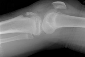
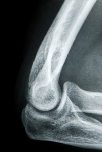
An X-ray of a knee joint affected by an avulsion fracture where the ligament tore a part of the bone away from the knee.

The April 2017 issue of the Journal of the American Academy of Orthopedic Surgeons features a compelling review article covering the topic of lower extremity avulsion fractures in pediatric and adolescent athletes. Drs. Jonathan Schiller, Steven DeFroda and Travis Blood discuss the effects of misdiagnosing lower extremity avulsion fractures. Avulsion fractures are more common in adolescents and happen when there is an injury to the bone in a place where a tendon or ligament is attached. The fracture causes the tendon or ligament to pull off a piece of bone.

Types of Avulsion Fractures

The JAAOS article discusses the most common places where the pediatric athlete will get avulsion fractures. The pediatric athlete has growth plates that are still open and attached to the tendon. Typically the growth plate is weak and when there is a traumatic event, the plate can often suffer an avulsion fracture. 

The most common places where pediatric patients will have avulsion fractures are at the anterior superior iliac spine and the anterior inferior iliac spine along with the ischial tuberosity. It is extremely important for medical professionals to conduct a plain x-ray when physically examining a patient in this case. The physical examination could be consistent with a muscle strain or apophysitis.  Apophysitis tend to occur with overuse whereas an avulsion fracture tends to happen acutely. The athlete may describe a popping sensation at the time of the injury.

Another avulsion fracture, tibial tubercle avulsion, is not very common, but requires immediate surgical fixation. A healthcare provider should be quite cognizant if their patient comes in with knee pain that could perhaps be the result of a tibial tubercle avulsion fracture.

How to Treat Avulsion Fractures

The most common way to diagnose an avulsion fracture is by x-ray. However, other injuries may require the use of a CT scan or MRI. CT imaging is more commonly used in the case of a tibial tubercle avulsion fracture. In general, treatment for this type of avulsion fracture is conservative unless there is a significant amount of displacement.

Nonsurgical treatment is divided up into a five stage protocol, which consists of:

  1. Ice, rest, anti-inflammatories for the first week of the injury  
  2. From week one to week three, involves gentle passive range of motion and assisted partial weightbearing is performed
  3. Resistance training occurs
  4. Allows aggressive stretching and sport specific activity after one to two months
  5. Two months post injury when the athletes are returned back to sports without restriction

Ischial Tuberosity

The ischial tuberosity is the origin of the hamstring muscles which involve the long head of the biceps femoris, semitendinosus and semimembranosus. Avulsion fractures in this area are caused by a forceful flexion of the hip joint and extension at the knee. It typically takes place during sprinting or kicking. If the avulsion fracture is displaced more than 15 mm, surgery is recommended.

Anterior Superior Iliac Spine 

These fractures are secondary to increased force from the sartorius and tensor fascia lata. They both originate from the anterior superior iliac spine. Nonsurgical management is recommended except if displacement is greater than 2 cm. These typically occur as a result of sudden straining, twisting, such as in baseball, jumping, and sprinting.

Anterior Inferior Iliac SpineAvulsion fracture of the spine

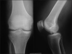
AIIS fractures are secondary to an increased force from the rectus femoris.  AIIS injuries typically occur during forceful hip hyper-extension and knee flexion. Conservative treatment is recommended for these avulsion fractures unless displacement is greater than 2 cm.

Pubic Synthesis and Iliac Crest 

These avulsion fractures are rare and occur from increased activity or force from adductor tendons, which originate from the pubic synthesis. These tend to occur during kicking type activity.  An MRI scan is typically needed to facilitate a diagnosis in this area. The rectus abdominis muscles attached to the iliac crest and avulsion fractures can occur during forceful turning or lateral flexion of the area. If the fracture is greater than 3 cm of displacement, surgery would be recommended.

Tibial Tubercle 

These fractures occur with forceful contraction of the quadriceps. It is important to quickly diagnose this particular injury because nonsurgical treatment is not only ineffective, but can also cause the lower extremity to become dysfunctional. X-rays are sufficient to make the initial diagnosis, but CT scans are able to better classify the extent of the injury. Because of the strong contraction of the quadriceps causing fracture displacement, these injuries rarely heal without surgical intervention.

Greater Trochanteric 

Fractures of the hip are rare in the pediatric population and are typically encountered in obese patients. This type occurs with the hip in flexion and with external rotation. Nonsurgical treatment is recommended for all trochanteric fractures. Surgery should be considered if there was significant displacement at the site of the fracture greater than 2 cm.

If you are feeling any significant pain in your joints, regardless of if you are an athlete or not, visit Dr. Stacie Grossfeld of Orthopaedic Specialists, PLLC in Louisville, KY. With the help of Dr. Grossfeld, you can prevent further damage to your ligaments, joints, and bones while starting a treatment plan to fix any existing damage. Contact Dr. Grossfeld here or by calling 502-212-2663 today!

Love Your Heart: 10 Ways to Keep Your Heart Healthy and Avoid Heart Disease

heart diseaseCoronary Heart Disease (CHD) is the most common type of heart disease to affect Americans and is also the type of heart disease that people are most likely to be talking about when discussing heart disease. CHD occurs when plaque builds up in the coronary arteries. Coronary arteries are the enlarged veins that move blood into the heart. When plaque builds up in these arteries, they narrow and less blood is able to travel to the heart. Narrowed arteries and less blood flow increase the risk of heart attack or chest pain which means it is very important to keep your heart healthy and avoid heart disease.

June is National Safety Month and at Orthopaedic Specialists, PLLC, we like to promote safety through healthy living. Below we’ve outlined ten ways that you can keep your heart healthy and avoid heart disease.

10 Ways to Keep Your Heart Healthy and Avoid Heart Disease

  1. Eat a Healthy Diet

    One of the most important steps to take towards keeping your heart healthy is eating a healthy diet. Once a healthy diet is established, all other steps to keeping a healthy heart come much easier. A heart healthy diet includes fruits and vegetables, whole grains that are a good source of fiber, and fish. Doctors recommend incorporating fish twice a week into a diet targeting heart health. What you eat is important, but so is how much you eat. Once you have established a healthy grocery list, research portion control and practice eating correct portions instead of overloading your plate.

  2. Live an Active Lifestyle

    Most people are ready to give excuses when faced with living an active lifestyle. From working overtime to having a family, it is easy to put exercise on the back burner and never take it off; however, an active lifestyle is not as big of a burden as most people think. Getting only 150 minutes (2 and a half hours) of aerobic exercise in every week constitutes as having an active lifestyle. Only 2 and a half hours a week of exercise and you’re sure to have a healthier heart for it.

  3. Maintain a Healthy Weight

    Over time, healthy eating and an active lifestyle will lead to you being able to maintain a healthy weight, but in the meantime it is important to understand what a healthy weight for your build and body type is, as well as, if you are there or if you have some work to do. Start by measuring your Body Mass Index (BMI) to see if you are at a health weight or not. If you aren’t, a 12 week weight loss program is the best place to start. Most come with meal plans that are health conscious and the bundled packages are often easy to follow.

  4. Quit Smoking (If You Do)

    Putting cigarettes down is the single, most important thing you can do to ensure heart health and overall health. Smoking cigarettes is the top cause of CHD and once you give up smoking cigarettes, your chances for developing heart disease drops by 50%. There are hundreds upon hundreds of free guides out there to help you quit smoking, but you are more likely to quit with assistance from the National Health Society or Smokefree.gov – the American equivalent. Click here to learn more about the National Health Society and their stop smoking services.

  5. Keep Low Cholesterol

    The main culprit behind high cholesterol is saturated fat. By cutting back on the amount of saturated fat that you consume in your diet, you will increase your heart health quickly. There are some very easy ways to lower your cholesterol including: picking leaner cuts of meat up at the grocery store and opting for 1% fat or skim milk instead of whole milk. Talking to a doctor about high cholesterol is a step that may need to be considered, especially if you struggle with lowering your cholesterol through changes in diet and exercise.

  6. Manage High Blood Pressure

    If you take all of the steps above then blood pressure shouldn’t be much of a problem; however, some people do struggle more than others with high blood pressure. If you have taken the steps above and have not been able to successfully lower your blood pressure, look at your caffeine consumption and consider cutting back on that. Not a caffeine drinker? Then it may be time to contact your doctor and discuss options in medication.

  7. Drink in Moderation

    In small amounts, alcohol can be beneficial to your health; however, in excess it is more likely to be detrimental than helpful. If you are drinking more than one 12 oz. beer, one 5 oz. glass of wine, or one 1 oz. pour of liquor a day – it is time to revisit what moderation means. Not only can alcohol have negative effects on your heart, but it can also raise your blood pressure and lower the effective properties of blood pressure medication.

  8. Manage Stress

    Stress is a huge factor that can negatively affect your heart health, but thankfully there are many ways to help manage stress. In this day and age, it is understandable that one can get overwhelmed time and time again; however, the time we live in is not an excuse. Manage your stress through exercise, deep breathing, making time for hobbies, discussing things that are bothering you, and going easy on yourself in tough times.

  9. Lower Sodium Intake

    Lowering sodium intake is a lot more difficult than not salting your dinner, but it is possible! Holding back on shaking salt over your dinner plate is the best place to start. It may take some time, but eventually your taste buds will get used to the taste of food without salt. Then you can stop using salt completely! Be aware of hidden salts in packaged foods. If there is more than 1.5 grams of salt per 100 gram servings then the food is considered “high in sodium” and should be avoided.

  10. Get 5-a-Day Every Day

    Getting your five-a-day every day is a fun and challenging way to promote your own heart health. The FDA recommends getting five separate servings of fruit and vegetables in your daily diet. This doesn’t mean making five vegetable sides with dinner or eating five apples a day. Find creative ways to incorporate fruits and vegetables into your healthy diet. Some ways to do this include: cutting a fresh piece of fruit over your fibrous morning cereal or adding a vegetable garnish into the pasta sauce that you’ve recently started making meatless.

From this article, it should be clear that heart health and overall health go hand in hand. If you look further into lowering your blood pressure or reducing your cholesterol, you’ll see that all medical advice starts with the same steps: eat a healthy diet, live an active lifestyle, maintain a healthy weight, and stop smoking. These steps are easy to take and easy to make into life practices with a little bit of determination and a lot of patience with yourself. Avoid Coronary Heart Disease and start now! There’s no better time to choose to practice a healthy lifestyle than National Health Month, this June.

If you are interested in learning more about how to live a healthy lifestyle that promotes heart health, contact Dr. Stacie Grossfeld at Orthopaedic Specialists, PLLC in Louisville, KY by calling 502-212-2663 or visiting her website today.

Tumbling, Trampoline and Acrobatic Gymnastics – Louisville Orthopedic on Gymnastics Injury

gymnastics injuryResearch led by Xavier Grapton (2013) and colleagues examined gymnastics injury information induced by tumbling, trampoline usage, and acrobatic gymnastics. Over a 5 year period, researchers looked at 357 traumatic events in acrobats and gymnastics injuries were identified based on the location of the gymnastics injury, the type of tissue injured, and causation.

Researchers found that gymnastic injuries related to acrobatic moves typically occur in lower limbs and often involve ligament related damage. Wrist injuries tend to be common among acrobatic athletes. Trampoline related injuries involved the knees or forearm while gymnastic injuries involving tumblers proved to be more ankle related. Researcher found landing-induced gymnastics injuries common across all activities and involved incorrect form, as well as, the landing surface.

In Conclusion

In conclusion, researchers found that the most significant injuries that occur in acrobatic sports are similar to artistic gymnastic injuries. Most injuries result from falling and incorrect or incomplete landings. Research documents clearly stated that landing surface also greatly impacts the possibility of injury in gymnasts and acrobats.

What is the Risk of Tearing Your ACL on Synthetic Surfaces?

The risk of tearing your ACL on synthetic surfaces has become a hot debate among physicians, coaches, and players.

With the level of competition improving and more year-round programs developing, synthetic surfaces are becoming the arena for many sports. In an American Journal of Sports Medicine article, published in July, 2015, Dr. George Balazs and his team looked at 963 ACL injuries. These injuries were reported on soccer or football fields. These reports were used to determine if synthetic surfaces increased the risk of tearing your ACL.

They stated that since the introduction of artificial turf in the 1960s, use has been a source of controversy. In trying to determine whether they are placing the athletes at increased risk, they looked at many factors. For one, artificial turf has largely been used as a secondary option to grass fields; however synthetic surfaces have increased durability, equate less cost, and present more consistent field conditions than natural grass. They also noted there’s been a steady increase in the use of artificial turf among  competition levels since the ’60s.

Over the past five decades, manufacturers have made significant changes to synthetic field surfaces. Some changes include: the fiber type used and its density, an increase in under-field padding, and the addition of rubberized soles to give the surface a more natural feel. All these changes have been made to ideally decrease field-related injuries.Tearing Your ACL

The authors noted that biomechanical studies have generally supported increased friction force on all types of artificial turf.

Theoretically this increases the risk of injury relative to natural grass. The potential elevated risk of tearing your ACL is a critical concern to athletes, coaches, team trainers, and physicians. The authors also stated that their study would evaluate data related to the effect plain surfaces had on the ACL. After reviewing journal articles, they found  there was an increased risk in American football players, but not in soccer players.

Part of the issue on evaluating whether artificial turf is the cause of lower extremity injuries is the result of multiple factors that may influence athlete injury rates. These include: field moisture, precipitation, and the temperature. Other factors like player position, types of play, period in the game, timing of the game within the season, legal or illegal play by an opponent, and type of shoe wear also affect study results.

The authors stated that there are so many different variables outside of just “artificial turf” that there were some tentative conclusions that could be drawn from this review article. They stated that earlier generations of artificial turfs (densely woven nylon fiber carpets placed on shock absorb with padding) appeared to have an increased risk of ACL injury. They also stated that there is an observed increased risk of ACL injury for football players on modern artificial turf (which is synthetic fibers with rubberized infill) and this seems to increase with higher levels of competition.

In Conclusion

There was no indication that an athlete participating in soccer had increased risk of ACL injury on modern artificial turf. The authors felt that more comprehensive studies looking at other factors needed to be completed to give a better answer as to whether artificial turf does increase the risk of tearing your ACL. But, based on the literature they reviewed in their analysis study, they noted that American football players are at increased risk while soccer players are not at increased risk for tearing their ACL based on turfs types. So good news for soccer players, but not the best news for football players!

If you are on a sports team and want to learn more about this study – call Dr. Stacie Grossfeld of Orthopaedic Specialists in Louisville, KY today at 502-212-2663. If you have suffered a tear in your ACL and are in need of orthopedic care, visit Orthopaedic Specialists today.

How fast can you run a mile? Find out on July 20 at the Parkway Mile!

The Louisville Sports Commission originally shared this information through an email. As a member of the Louisville Sports Commission board, Dr. Stacie Grossfeld is sharing this information on her personal website.

Registration is now open for Louisville’s newest summer race, the Louisville Active Parkway Mile.

parkway mile

We’ve all trained and run 5Ks, half marathons, and some even triathlons. But have you ever wondered how fast your time would be if you ran just mile? Now you can find out. The Louisville Sports Commission has created the Louisville Active Parkway Mile, a fun one-mile run/walk taking place on Southern Parkway.

Beginning at 6:30 p.m. on Thursday, July 20, participants can take off from Southern Parkway and Woodlawn Avenue and take a straight and fast route down Southern Parkway to the finish line outside of Olmsted Academy South.

Parkway Mile Heats Include:

  • Elite Male (must meet qualifying standard of 5:00 mile)
  • Elite Female (must meet qualifying standard of 6:00 mile)
  • Open Male
  • Open Female
  • Masters Male (“Master” is anyone 40 years old and up, per USATF.)
  • Masters Female
  • Family Fun Mile (Not timed.)
  • High School Male
  • High School Female
  • Youth Mile (“Youth” are kids in middle school and younger.)

All heats, excluding the family fun mile, will be professionally timed. Awards will be presented to the fastest, and all will receive post-race refreshments including Powerade, water, fruit, and other goodies.

Register today for only $20 to participate in Louisville’s newest and fastest race! Visit https://runsignup.com/Race/KY/Louisville/ParkwayMile to find out more information and sign up. Also follow us on Facebook for race updates at https://www.facebook.com/ParkwayMile/.

Ulnar Collateral Ligament Injuries: An Overview

ulnar collateral ligament injuriesThe human elbow is an intricate part of the body. While it is essentially a hinge joint and looks fairly straightforward – the elbow is made up of a complicated series of bones, joints, and ligaments. Within the elbow the radius and ulna must rotate and slide with the humerus to bend or straighten the arm. These movements are possible with the help of ligaments and there are two main ligaments that are important in order to achieve optimal elbow maneuverability: the lateral collateral ligament and the ulnar collateral ligament.
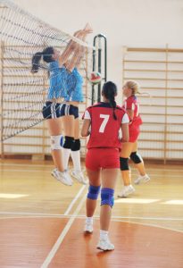
Both ligaments work together to keep the humerus connected to the ulna and to keep the ulna in place. Elbow stability depends on these two ligaments and when one is injured, it seriously affects how the elbow functions. Often, the ulnar collateral ligament is hurt through overuse or repetitive use. Not surprisingly, athletes who often depend on throwing motions are most likely to report ulnar collateral ligament injuries. This especially applies to baseball pitchers, quarterbacks in football, and softball players in the outfield. There are three phases to ulnar collateral ligament injuries and being aware of the stages can help to prevent further damage from occurring.

3 Phases to Ulnar Collateral Ligament Injuries

Phase 1: Microtearing

Microtearing is the first phase to a full blown ulnar collateral ligament injury. If it can be prevented then the injury has a better chance of being prevented. The small tears through the ligament occur when the force that the tissues are handling is heaving than the strength of the ligament. When the ligament starts to tear, it is not extremely noticeable. Often it takes months or years of repetitively hard throwing motions to begin the microtearing process.

Phase 2: Degeneration

Degeneration occurs after microtearing when the build-up of minute tears starts to wear away at the ulnar collateral ligament and leaves lasting damage that the body cannot keep up with repairing. When degeneration sets in, pain may start to occur in the elbow and swelling is often seen from the outside. If ulnar collateral ligament injuries are detected in this phase, the healing process can remain nonsurgical. Often, even when ulnar collateral ligament injuries have reached the degenerative stage, rest and modification of activity can stop the injury from worsening and allow the body a chance to start healing.

Phase 3: Rupturing

When an ulnar collateral ligament ruptures, it is accompanied by a loud pop and immediate pain. When an ulnar collateral ligament ruptures, the elbow will lose most of its stability. Sometimes a rupture can occur from just one powerful ligament tearing throw by an athlete; however, more often, the ligament will rupture after years of micro tears and degeneration has already weakened the ligament. When a rupture occurs, surgery is the main option for recovery and a board certified orthopedic surgeon can tell if an ulnar collateral ligament rupture will require repair or reconstruction.

Athletes who often use their arms to pitch, throw, or launch repetitively should be aware of the three phases that lead to ulnar collateral ligament injuries and seek qualified medical attention if they start to feel any of the following symptoms:

  • Pain throughout the inside (side closest to your body) of the elbow
  • Any popping sounds or grinding noises
  • Swollen areas or bruising along the inside of the elbow

A simple MRI can detect any potential ligament injuries. If ulnar collateral ligament injuries are detected an orthopedic physician can help to set up a treatment plan. However, if the damage is too far along then surgery may be the only option.

If you are feeling any significant pain in either of your elbows, regardless of if you are an athlete or not, visit Dr. Stacie Grossfeld of Orthopaedic Specialists, PLLC in Louisville, KY. With the help of Dr. Grossfeld, you can prevent further damage to your ligaments, joints, and bones while starting a treatment plan to fix any existing damage. Contact Dr. Grossfeld here or by calling 502-212-2663 today!

Why Do Female Athletes Tear Their ACL at a Higher Rate Than Male Athletes?

There is a lot of debate over the causes of ACL tears among women and the difference when compared to male athletes. It is proven that women tear their ACLs at a higher rate than men; however, the cause has not be pinned down. While the cause hasn’t been determined yet, there are many theories surrounding this type of injury and why it is so frequent among female athletes.

Dr. Grossfeld explains ACL tears in girlsThe Theory

One theory focuses on the fact that as girls go through puberty, they develop strength differences in their hamstrings versus the quads. This change effects the way females land from a jump and causes some loss of hip strength. This theory has inspired many scientific studies including one in May of 2016. In May, the American Journal of Sports Medicine published a study from Dr. Catherine Wiles – out of Australia – where she specifically looked at how young girls changed their landing technique throughout the adolescent growth spurt.

The study was very unique in the sense that Dr. Wiles followed the girls as they went through puberty and measured then studied the way their lower extremities matured during this time period. Dr. Wiles measured ground reaction forces, lower limb muscle activity, and kinematic data while the girls performed horizontal leaps. The authors noted that during the adolescent growth spurt, girls experienced a vast number of rapid insightful changes to the musculoskeletal system including the structure and the function.

The Findings

Through puberty, there is an increase in height and lower limb length as well as increases in anterior knee laxity. There is a lack of hamstring strength development that is relative to the strength of quadriceps. After four different test periods, Dr. Wiles and researchers found that over the course of puberty Test #1 produced completely different results compared to Test #4. This is in reference to the less flexion that the girls had in their knee when they landed their horizontal jump. They found that as the girls went through maturity, they displayed a greater hip external rotation, also.

When specifically looking at the knee, the researchers determined that the girls experienced a decrease in external rotation moment as they matured. It is possible that increased knee flexion acts as a landing strategy to protect the internal structures of the knee. This then decreases the risk of ACL injuries. A knee flexion that goes less than 22 degrees during landing can increase the potential that an athlete’s quadriceps will strain the ACL. This then increases the likelihood of an ACL rupture.

They found that with an isolated quadricep load of 200 Newtons in ACL loads with the highest at 45 Newtons at 15 degrees of knee flexion that the force on the ACL decreased by approximately 10 to 15 Newton’s with every 15 degree increase in knee flexion. This means that researchers found female athletes with a higher amount of knee flexion during a horizontal jump produce less force on the ACL.

However, as the girls matured in this study, the amount of knee flexion reduced as they did the horizontal jump. This could explain why female athletes are more prone to ACL injuries than their male counterparts.

Healthy Bones: Osteoporosis

Bone Health and Osteoporosis 
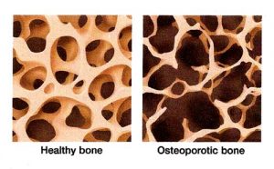
This month we are focusing on bone health. Unless you have recently sustained an injury to your bones, then it’s unlikely you’ve been putting much thought into their health. Since we can’t see our bones, it is much more difficult to remain aware and alert to their needs. For example, osteoporosis, which is a condition that degrades bones, is a common disease and yet most people aren’t aware of its serious effects on bone health.

Effects of Osteoporosis on Bone Health 

According to the National Osteoporosis Foundation, about 54 million Americans have osteoporosis. Approximately one in two women and up to one in four men age 50 and older will break a bone because of osteoporosis. The condition typically appears in women when they start menopause at age 50-55 . Male osteoporosis tends to occur after age 70. The start of osteoporosis is related to a reduction of the hormones estrogen and testosterone.

Osteoporosis is a silent disease. The first sign of the condition may only appear after a fragility fracture has already occurred. These kinds of fractures are most common in the wrist, spine, and hip. Studies show that someone who has had a fractured hip due to bone density decrease has a much higher risk of fatality because of the injury than someone else with the same fracture unrelated to osteoporosis.

Other than broken bones, osteoporosis can cause height loss or curvature of the spine. If you are experiencing either of those symptoms, it could be wise to get in contact with a healthcare professional.

Prevention

Prevention is the best form of treatment of osteoporosis. A few ways you can work to prevent the condition is by incorporating more calcium and vitamin D supplements into your diet. Some foods rich in calcium include dark leafy greens, cheese, yogurt, broccoli, and almonds. Sunlight is the vitamin D source most people know, but there are also other ways to incorporate vitamin D into your life even if you can’t get outside for lengthy periods of time. Foods such as salmon and other fatty fish, egg yolks, and portobello mushrooms are also good sources of vitamin D.

Other forms of prevention include frequent exercise, reducing caffeine and alcohol intake, as well as eliminating any smoking habits. These activities not only help prevent osteoporosis but also help to improve general bone health.

If you think you may be at risk for osteoporosis, there is also a bone density screening you can opt for. It is called a DEXA scan and is somewhat similar to an x-ray.

Treatment

There is no explicit cure for osteoporosis, however there are medications that can help reduce loss of bone density. Calcium and vitamin D supplementation can only go so far to help prevent the onset of osteoporosis, but they cannot help to reverse the effects of the condition. Only the group of FDA approved medications for treatment of osteoporosis can be used to rebuild bone mass such as: Prolia  and Forteo. Only an FDA approved medication for treatment of osteoporosis can improve and rebuild the bone density and strength. Ask your healthcare provider which agent they recommend before you try to self-prescribe.

Subchondroplasty or SCP for Bone Marrow Edema or Insufficiency Fractures

subchondroplastyMany people suffer from bone marrow edema or insufficiency fractures. And the long term effects of these conditions can be serious. Edema and insufficiency fractures can injure a person’s bone marrow and cause a long list of medical complications.

Fortunately, there are a variety of medical treatments available to treat bone marrow edema and insufficiency fractures. People with the most chronic and severe cases may benefit from subchondroplasty, a treatment provided by a board certified orthopedic surgeon like Dr. Stacie Grossfeld. Subchondroplasty can improve a person’s pain and also decrease the number of physical limitations often associated with bone marrow edema or insufficiency fractures.

What are Insufficiency Fractures?

Insufficiency fractures are a type of stress fracture. They happen due to normal stress on a bone that is weak. Insufficiency fractures are more common in older people, and particularly in older women.

What Causes Bone Marrow Edemas?

When you suffer from edema, your blood cells go to the injured site, causing a pooling of fluids. Injuries such as fractures or broken bones result in bone marrow edema. They may also be the result of ligament injuries, bruising on bones or inflammation. Osteoarthritis and osteoporosis are regularly associated with bone marrow edema. Bone marrow edemas can negatively impact a person’s overall health. If you are suffering from this condition, seeking treatment is very important.

An Overview of Subchondroplasty as a Treatment for Edema

If you are experiencing edema, and you have not gotten better from medications and rest, you may benefit from subchondroplasty. Subchondroplasty, also known as SCP, is a procedure done for patients that have insufficiency fractures or bone marrow edema. Typically it occurs in patients with osteoarthritis. An MRI scan diagnoses it.

The surgical procedure is an outpatient procedure that takes about 20 minutes to complete. It involves a knee arthroscopy where a high definition television camera is inserted into the knee joint. A trocar, which is like a large needle, is injected into the area where bone marrow edema was identified on the preoperative MRI scan. Then a special type of bone graft is injected. The bone graft instantly heals the bone marrow edema or insufficiency fracture.

What to Expect after Subchondroplasty?

Following subchondroplasty, patients are able to bear weight immediately. And generally within 24 to 36 hours, people also experience a noticeable reduction in pain. Dr. Grossfeld has been performing subchondroplasty for about a year and while doing so has had excellent results. For more information and to schedule an appointment with Louisville orthopedic Dr. Stacie Grossfeld, call 502-212-2663.

Dr. Stacie Grossfeld of Louisville’s Orthopaedic Specialists PLLC Named 2017 Volunteer of the Year by the YMCA at Norton Commons

ymca at norton commons

Dr. Grossfeld accepts her award for the 2017 Volunteer of the Year from Mike Bramer, the District Executive Director at the YMCA at Norton Commons.

Louisville, KY – April 27, 2017 – Dr. Stacie Grossfeld, orthopedic surgeon at Orthopaedic Specialists, PLLC, has been named Volunteer of the Year by the YMCA at Norton Commons in Louisville, Kentucky. Since 2015, Dr. Stacie Grossfeld has served on the Board of Directors for the organization, and has regularly connected the YMCA with potential business partners since joining.

Since joining the Norton Commons YMCA Board of Directors, Dr. Stacie Grossfeld has been happy to help fundraise for the Y at Norton Commons.

Some examples of Dr. Grossfeld’s involvement with the YMCA at Norton Commons include:

  • Finding and giving the Y a $4,000 blood pressure machine for members to use
  • Securing donations and contributions to the YMCA at Norton Commons
  • Encouraging individuals and families to join the YMCA through her orthopedic and sports medicine practice

Describing their experience with Dr. Grossfeld, leaders at the YMCA at Norton Commons explain: “Stacie is a tremendous asset to the YMCA at Norton Commons and she is very deserving of our Volunteer of the Year Award!”

The YMCA at Norton Commons is one of the newest YMCA facilities to open in the Greater Louisville area. At the Norton Commons YMCA, families can take advantage of a wide variety of programs including:

  • Nutritional Consultation
  • Personal Training
  • Youth Fitness Fundamentals
  • YMCA Weight Loss Challenges
  • Fundraisers through the YMCA Teen Leaders Program

The YMCA at Norton Commons also offers a warm water pool for recreational activities, a lap pool for exercise, a sauna, spa, group exercise and cycling classes, a fitness center, supervised Kid’s Club, Club Y for preteens, a full gymnasium, and more.

To read more about how Dr. Stacie Grossfeld helps give back to her Louisville, KY community and what the YMCA at Norton Commons has to offer to children and adults alike, click here.