When your doctor needs to see what’s going on inside your body, they’ll likely order a medical imaging test for you. Medical imaging tests, such as MRIs, X-rays, and CT scans, use different kinds of energy to provide detailed pictures of the inside of your body. In doing so, the imaging tests can help experienced medical professionals diagnose injury and disease, provide treatment options, and monitor existing conditions.
At some point, almost everyone needs some type of medical imaging test. When the time comes, your doctor will provide you with information, but you’ll still likely have questions about the procedure or the differences between the modalities. That’s why we’ve broken down three common tests in the paragraphs below.
An Overview Of 3 Common Medical Imaging Tests
MRI
Magnetic Resonance Imaging (MRI) tests use a magnetic field and computer-generated radio waves to produce high-resolution images of your organs, bones, tissues, and other body structures. A technician will monitor you as you lay down in the MRI machine, which is a long, narrow tube. Usually taking between 30-60 minutes, the process is non-invasive and painless.
MRI tests generally provide more comprehensive, clearer images than CT scans and x-rays, but are also more expensive and time-consuming. Doctors often choose MRIs are to scan the brain, spinal cord, and nervous system, as well as muscles, tendons, and ligaments. Doctors can detect some conditions only with an MRI. These include inflammation, certain illnesses, and types of cancer. Due to the magnetic field, MRIs should not be used by people with metal implants, such as a pacemaker, cochlear implant, or implanted insulin pump.
X-Ray
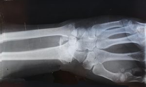
X-ray tests produce images of bones in particular. These tests work by passing x-ray beams through your body that are absorbed in different amounts by different mediums. Denser materials, like bone, show up white, while less dense structures such as muscles and fat show up gray. Compared to MRIs, x-rays are faster, easier, and cheaper—but also may show less detail. X-rays also utilize low-dose radiation, which is not harmful, but can be a concern for some patients.
X-rays are best used to display bone conditions, including arthritis, bone fractures, cavities in teeth, osteoporosis, and bone cancer. Doctors can also use them to diagnose chest conditions such as lung infections, congestive heart failure, and breast cancer.
CT Scan
A computerized tomography (CT) scan takes a sequence of x-ray images around your body, then uses a computer to create a comprehensive cross-sectional image. A CT scan is more detailed than an x-ray, but not as much as an MRI. However, a CT is much faster and more affordable than an MRI. Sometimes it is a good option for victims of car accidents or internal trauma.
CT scans are excellent at showing skeletal system problems, bone mineral density, blood vessels, lung issues, and abdominal abnormalities. They are also good for monitoring tumors and cancer treatment over time.
If you have a condition that you think requires medical imaging, contact your physician for more information. Dr. Stacie Grossfeld at Orthopaedic Specialists PLLC serves people of all ages throughout the Louisville, Kentucky-region in need of all types of orthopedic treatment and sports medicine. For more information or to schedule an appointment, call 502-212-2663 today.
Recent Comments