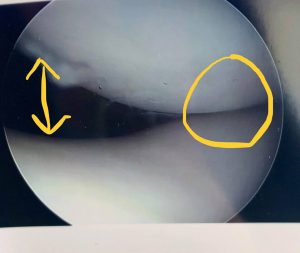
This is a condition where your kneecap is tilted and compresses on the femoral groove abnormally. Imaged below is a picture of a kneecap joint with lateral patellar compression syndrome. You can see where on the right side the bones are touching each other and on the left side there’s a big open space. There should be an even space on both sides.

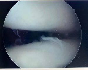
This picture shows how the kneecap looks after I have performed a lateral release. This should give the patient almost immediate pain relief. This is a diagnosis sometimes difficult to figure out because there is not a great imaging modality to document. Many times it is more of a dynamic problem and cannot be statically imaged.


Recent Comments