My 12 year old son, poo poos it because we don’t have 4K monitors in the operating room and it’s only 1080p, however in 1080p this is what knee osteoarthritis looks like:
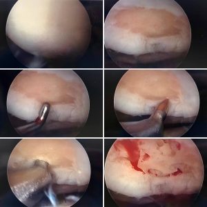
The first image (upper left ) is a normal knee with normal white, smooth cartilage covering the end of the femur bone. Image number two at the top right reveals a large arthritic area which is the pale orange color surrounded by the white articular cartilage. The pale orange area is the exposed bone with the cartilage missing (the definition osteoarthritis). The middle row left is my metal probe in the knee joint pointing at the area of osteoarthritis.
Middle row right reveals a chondro-pick, which has a sharp pointed tip. Yikes!!! We use this medieval looking instrument to literally poke holes in the area of exposed bone. Which I am doing in the bottom left image. Why??? Penetrating the exposed bone causes the bone to bleed. That will attract healthy cells which in turn forms fibro-cartilage. This scar tissue type cartilage will fill in the area of exposed bone reversing the process. In the last image, lower right, you can see the bone bleeding. Mission accomplished.
Six weeks with no weight bearing on that leg while the new cartilage is generated. Think of it like planting new grass seed, you can’t walk on it while it is growing, or it will be destroyed. In six weeks, happy knee and happy patient!
Recent Comments