

Diabetes and Surgery: Why You Should Ask Your Patients if They are Diabetic Before Scheduling Surgery

Person taking A1c Blood Glucose test

Diabetes is a condition that tends to affect the daily life of individuals. However, it goes beyond that. Diabetes can alter surgical plans if HbA1c test levels are too high, which is why primary care doctors should be asking patients to take frequent tests before surgery.

Key Takeaways

  • If the results of an HbA1c test are over 8%, surgeries will need to be canceled because of anesthetic complications and a higher risk of post-operative complication.
  • If the Hemoglobin A1C is 7% or lower, surgeries will likely be green-lighted.
  • HbA1c test levels can often be lowered by implementing lifestyle changes like consistent exercise, eating whole foods, and managing stress or by medication.

When is it Safe to Operate with Diabetes?

An A1c test shows your average level of blood glucose (blood sugar) over a time period of approximately 2-3 months. Before a surgery is scheduled and before it takes place, doctors should be either asking when a diabetic patient’s last HbA1c test was or asking the patient to take a test for accurate readings.

In general, surgeries will not be scheduled or completed if an A1c test result is over 8%. This puts diabetic patients at a higher risk of anesthetic or post-operative complications. A1c results of 7% or lower are ideal for a safe surgery.

What Are the Risks of Operating with a High A1c?

Diabetic patients with a blood glucose level of 8% or higher are more at risk of:

  • Infection
  • Poor wound healing
  • Longer recovery periods
  • Hospital readmission
  • Hyperglycemia or hypoglycemia
  • Electrolyte imbalance
  • Diabetic ketoacidosis

It is very important not only to monitor your glucose levels before surgery, but afterwards as well.

How to Lower Your A1c

Sometimes, diabetic patients will receive medication to help lower blood glucose levels. However, lifestyle changes are more often the suggestion. Your doctor can help you prepare for surgery by monitoring lifestyle elements like:

  1. Diet. Committing to whole foods like vegetables, whole grains, legumes, nuts, lean protein, and fruit is one of the most common and successful ways to manage blood glucose. You may even be referred to a nutritionist to help you achieve a sustainable diet.
  2. Exercise. Another highly successful blood glucose management tool is movement. It doesn’t have to be high-intensity or long workouts, but consistency is key. Taking walks, joining a low-impact exercise class at a gym, or hiring a personal trainer to help meet your goals is one of the best ways to lower HbA1c levels.
  3. Stress Management. High levels of stress may contribute to high levels of blood glucose. Having stress management strategies like yoga, meditation, and other hobbies that you enjoy increase mental health, which in turn has an impact on physical health.
  4. Curbing Smoking Habits. Nicotine has proven to increase blood glucose levels, increase the risk of developing diabetes, interfere with the effectiveness of insulin, and makes it harder to manage diabetes.

Frequently Asked Questions about Diabetes and Surgery

1. Does diabetes increase my risk of complications from surgery?

Yes, diabetes can increase complications and risks of surgery. Elevated blood sugar before, during, and after surgery is linked to slower wound healing, a higher risk of infection, and longer hospital stays. Diabetes can also affect the heart, kidneys, and nerves, which matter for anesthesia and recovery.

2. What should I do with my diabetes medications and insulin before surgery?

This depends on the specific medication and the type of surgery, so you should follow the instructions from your surgeon, anesthesiologist, or diabetes provider rather than adjusting things on your own. For example, certain drugs (such as SGLT2 inhibitors) are commonly paused several days beforehand, while others may be continued or adjusted. Insulin doses are often reduced, particularly long-acting insulin the night before, because you’ll be fasting. Ask for written instructions in advance and bring a list of everything you take.

3. How will my blood sugar be managed during and after surgery?

Your blood sugar is typically checked frequently on the day of surgery, and the team may use insulin (sometimes through an IV) to keep it within a target range. Fasting, stress, and anesthesia can all push glucose up or down, so close monitoring is necessary. After surgery, you’ll usually continue to be monitored until you’re eating normally and can return to your regular routine.

If you or someone you love needs an orthopardic surgeon in the Louisville, Kentucky-area, board certified sports medicine physician Dr. Stacie Grossfeld at Orthopaedic Specialists PLLC can help. Orthopaedic Specialists PLLC is accepting new patients, and same day appointments are available. For additional information or to schedule an appointment, please contact Orthopaedic Specialists PLLC today at 502-212-2663.
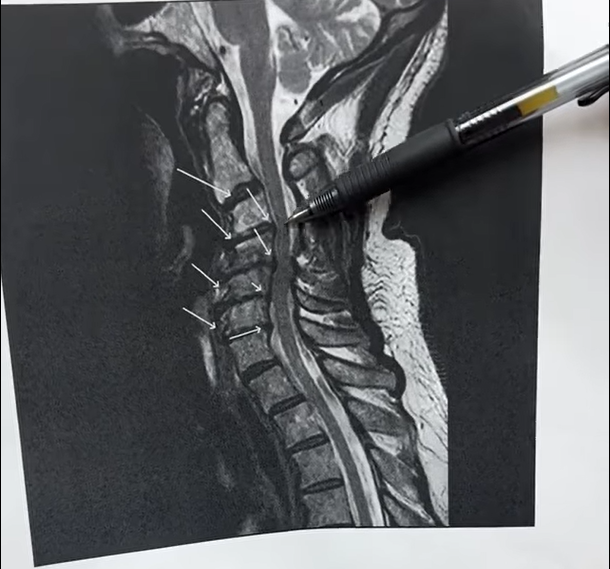
Neck Pain and the Spine: What Does an Abnormal Cervical MRI Look Like?

Cervical Spine MRI Abnormalities that Cause Neck Pain

Taking a look at an abnormal cervical and neck MRI, Dr. Grossfeld points out where to look for abnormalities. Abnormalities in the cervical region of the spine can cause serious neck pain. Knowing where and what to look for can help doctors diagnose where the pain originates from.

Key Takeaways:

  • Abnormalities in the cervical spine are commonly found in the vertebral bodies and vertebral discs.
  • Normal vertebral bodies look square and do not push or bulge into the cerebrospinal fluid or the spinal cord itself.
  • Normal vertebral discs look homogenous in both color and size.

Where to Look for Cervical Abnormalities

In most cases, cervical abnormalities will be found in either the vertebral discs or the vertebral bodies. On an MRI, normal-looking vertebral bodies will be square and positioned outside of the spinal cord and cerebrospinal fluid. Normal-looking vertebral discs will be dark and homogenous in size.

Vertebral bodies are the individual bones of the vertebral column that are cushioned by the vertebral discs, which are made of cartilage in two parts: annulus fibrosus (the outer portion) and nucleus pulposus (the inner core). They make up a majority of the spine, which protects the spinal cord and connecting nerves.

However, if they interact with each other and the cerebrospinal fluid and spinal cord in the wrong way, they can cause a lot of pain. Some of the most common causes of abnormalities of these two parts of the spine include:

  • Disc Degeneration
  • Herniated disc
  • Cervical stenosis
  • Osteoarthritis
  • Cervical Spinal Cord Compression

How to Treat Neck Pain at Home

Neck pain can be caused by trauma or degenerative conditions, which are treated differently. If you have been hurt or are experiencing long-lasting neck pain, see a doctor for a diagnosis and unique treatment plan. However, most conservative neck pain treatment plans may include:

  • Over-the-counter pain medications
  • Physical therapy to strengthen neck, shoulder, and back muscles and improve flexibility
  • TENS electrical nerve stimulation therapy
  • Steroid injections to relieve inflammation
  • Heat therapy
  • Massage therapy

How to Prevent Neck Pain

Sometimes, neck pain due to cervical spine abnormalities appear due to factors out of our control, such as age or degenerative conditions. But, to help protect yourself from factors within your control:

  • Strengthen and exercise the muscles in your neck, upper back, and core. This can help to protect your spine and is important for maintaining good posture.
  • Practice healthy sleep habits. Invest in a supportive pillow that is made for your preferred sleeping position and try to keep your body aligned as your sleep.
  • Stretch often. Making a habit of stretching keeps muscles and tendons from becoming inflexible and vulnerable to injury.
  • Avoid carrying heavy weight on your shoulders frequently. Backpacks, suitcases, boxes, and other equipment are better off carried on wheels or with someone else to mitigate risk of injury.

If you or someone you love has suffered a neck injury in the Louisville, Kentucky-area, board certified sports medicine physician Dr. Stacie Grossfeld at Orthopaedic Specialists PLLC can help. Orthopaedic Specialists PLLC is accepting new patients, and same day appointments are available. For additional information or to schedule an appointment, please contact Orthopaedic Specialists PLLC today at 502-212-2663.
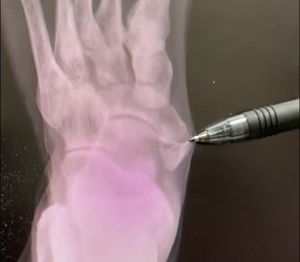
What is an Accessory Navicular?

Accessory Navicular Bone on Xray

An accessory navicular is an extra bone that people can be born with in their foot or feet. It is located on the inner side of the foot right above the arch, and gets its name because it grows so near the primary navicular bone. It also goes by its Latin names, os naviculare or os tibiale externum.

Key Takeaways:

  • Accessory navicular bones are congenital, meaning that they are present at birth.
  • An accessory navicular bone that causes pain or other symptoms is known as accessory navicular syndrome.
  • It is very rare to have to remove an accessory navicular bone because of accessory navicular syndrome.

What Causes Accessory Navicular Bones?

There is no real distinguishable cause of accessory navicular bones, though research shows that they can be genetic. They are congenital, meaning that they are present at birth and not something developed later in life. These extra bones form when babies develop a skeleton in the womb. All bones start out as cartilage, and if there is extra cartilage in the foot, then bone tissue will develop around it and create an extra bone.

There are three types of ways an accessory navicular bone may develop (Cleveland Clinic, Accessory Navicular Bone):

  1. Type 1: the accessory bone is embedded into the tendon that connects your calf to the inner side of your foot and ankle (posterior tibial tendon). These are small and are unlikely to cause symptoms.
  2. Type 2: the accessory bone(s) grow next to the navicular bone and connect via extra cartilage. These are larger than type 1 and are more likely to cause symptoms.
  3. Type 3: the accessory bone is fused to the primary navicular bone. It may cause symptoms if it comes into contact with tendons or nerves.

What is Accessory Navicular Bone Syndrome?

In many cases where they are present, an accessory navicular bone(s) is asymptomatic. Many people may not even know that they have an extra bone. However, if it causes pain or other symptoms, then there may be a diagnosis for Accessory Navicular Bone Syndrome.

This condition occurs most often around puberty, when the bones start maturing more (Foot Health Facts, Accessory Navicular Syndrome). It can happen on its own, but injuries and habits like wearing tight shoes or overusing your feet and ankles can contribute to it. It is characterized by symptoms like:

  • A visible bony prominence on the inner foot above the arch
  • Redness, pain, throbbing, and swelling on the inner foot above the arch, especially after activity
  • Limping
  • Pain while standing

How to Treat Accessory Navicular Bones

Most cases of Accessory Navicular Bone Syndrome are successfully treated with conservative methods. This includes:

  • The RICE method
  • Over the counter pain medication
  • Medical devices like a boot, crutches, or a cast to immobilize and shift weight during the healing process
  • Physical therapy
  • Shoe inserts

It is very rare that an accessory navicular bone will need to be surgically removed. If surgical intervention is necessary, though, a doctor will go in, remove the bone, then reattach any tendons that may have previously been attached to the accessory navicular.

If you or someone you love has suffered a foot injury in the Louisville, Kentucky-area, board certified sports medicine physician Dr. Stacie Grossfeld at Orthopaedic Specialists PLLC can help. Orthopaedic Specialists PLLC is accepting new patients, and same day appointments are available. For additional information or to schedule an appointment, please contact Orthopaedic Specialists PLLC today at 502-212-2663.
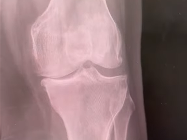
Bone on Bone Arthritis and the Stages of Osteoarthritis

bone on bone arthritis shown on an xray

Bone on bone arthritis is a common term that describes end-stage arthritis. When arthritis reaches “end-stage,” there is no articular cartilage left in the joint space to cushion the bones, which causes them to grind together and get the name bone on bone arthritis.

Key Takeaways:

  • Osteoarthritis is categorized by four stages: early, mild, moderate, and severe.
  • End-stage arthritis does not always mean surgery or knee replacements.
  • Joints with end-stage arthritis may still respond positively to conservative, non-surgical care like physical therapy, corticosteroid injections, and activity modification.

What are the Stages of Arthritis?

Since bone on bone arthritis is end-stage, that means that there are other stages of osteoarthritis as well. This is true! Osteoarthritis is often separated into 4 sections: early, mild, moderate, and severe (What are the 4 stages of osteoarthritis?, Geisinger).

1. Early Stage Osteoarthritis

This stage goes undetected often because the symptoms are negligible and physical changes in the joint can be undetectable on imaging tests. Stiffness after a joint has been resting in the same position for a while or soreness after physical activity may be indicators of early stage arthritis.

If arthritis is caught this early, a lot can be done to slow the progression of the condition. Lifestyle changes like an increase in activity, change in diet, or swap to low-impact physical activities like swimming, walking, or biking are all common.

2. Mild Stage Osteoarthritis

If arthritis enters the mild stage, the indicator symptoms will likely grow stronger and more frequent. In addition, visible changes in the joint may start showing up in imaging tests, such as bone spurs or cartilage damage. Consistent morning stiffness and pain or swelling during activity may occur and cause daily activities like opening jars or climbing stairs to become more difficult.

At this point, patients are still advised conservative treatment. Heat therapy, ice therapy, physical therapy, and supportive devices like sleeves or shoe inserts are all recommended. Over-the-counter pain medication may also be suggested, but you should talk to your doctor before taking anything consistently and for long periods of time.

3. Moderate Stage Osteoarthritis

By the third stage, symptoms are often persistent and affect daily activities with pain, stiffness, swelling, and fatigue. Stiffness may not always fade, and swelling can interfere with range of motion for several joints. Imaging tests should be able to pick up on these changes if symptoms grow to be this noticeable. You’ll likely see narrower cartilage space between bones in the joint, and your bone density may increase.

In addition to the non-invasive treatments like lifestyle changes, support devices, and physical therapy, your doctor may also prescribe corticosteroid or hyaluronic acid injections to help manage pain. Occupational therapy can address any changes in motion and help you adjust to new ways of moving as well.

4. Severe Stage Osteoarthritis

Stage four arthritis, also known as severe arthritis, end-stage arthritis, or bone-on-bone arthritis, often present a lot of issues for individuals. With extensively worn cartilage, prominent bone spurs, and extreme stiffness, swelling, or deformity in joints, it can greatly affect quality of life. If conservative treatments do not work, your doctor may suggest joint replacement surgery.

However, just because you see bone on bone arthritis on your imaging tests, it doesn’t automatically mean you should have surgery. If you are responding well to conservative treatments and are feeling good, surgery should still be a last-resort. Not every osteoarthritis case is the same, so not every treatment path is either!

If you or someone you love has suffers from osteoarthritis in the Louisville, Kentucky-area, board certified sports medicine physician Dr. Stacie Grossfeld at Orthopaedic Specialists PLLC can help. Orthopaedic Specialists PLLC is accepting new patients, and same day appointments are available. For additional information or to schedule an appointment, please contact Orthopaedic Specialists PLLC today at 502-212-2663.
Why Would I Need an EMG Nerve Conduction Study?

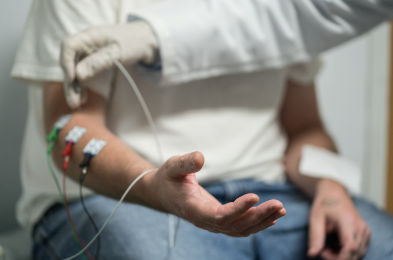
EMG nerve conduction study for cubital tunnel syndrome

Your doctor may order an electromyography test, also known as an EMG nerve conduction study, if they expect that you have a nerve injury such as carpal tunnel syndrome, cubital tunnel syndrome, or a nerve laceration.

Key Takeaways:

  • An EMG nerve conduction study is a test that checks how well your muscles and nerves are communicating with each other and if they are responding correctly.
  • Common symptoms that might necessitate an EMG nerve conduction study include muscle weakness, tingling sensations in your extremities, muscle spasms, and paralysis.
  • Carpal tunnel syndrome and cubital tunnel syndrome are both conditions that originate from pressure, inflammation, or injury to a nerve.

What is an EMG Nerve Conduction Study?

An EMG nerve conduction study tests the electrical signals your muscles make when they are at rest and when they are in use (Electromyography (EMG) and Nerve Conduction Studies, MedlinePlus). If they are responding correctly, a resting muscle will have no electrical activity and an in-use muscle will have consistent electrical signals that grow with the intensity of the movement.

What Should I Expect From An EMG Test?

During an EMG test, a doctor will insert a small needle with an electrode into the affected muscle, which is what records the electrical activity. Then, you will be guided to complete different movements, rests, and contractions with that muscle. The recorded readings will help get a clearer image of the muscle to nerve connection (EMG (Electromyography), Cleveland Clinic).

Your doctor also might utilize sticky electrodes to your skin above an affected nerve. Then, those electrodes are stimulated with small pulses of electricity, which in turn stimulate your nerves. The time it takes for your muscle to respond to the nerve will be recorded for analyzation.

What Symptoms and Conditions is an EMG Nerve Conduction Study Used for?

The most common symptoms that prompt an EMG include:

  • Muscle weakness
  • Tingling and/or numbness in your arms, legs, hands, feet, or face
  • Muscle cramps
  • Muscle spasms or twitches
  • Partial or full paralysis of any muscle

Common conditions that cause these symptoms are:

  • Carpal tunnel syndrome: Tingling and weakness in the fingers and hand due to a compressed median nerve.
  • Cubital tunnel syndrome: “Funny bone” pain that occurs due to inflammation or irritation of the ulnar nerve near the elbow.
  • Herniated disks: Back pain caused by the disks of your spine bulging out of place and putting pressure on the spinal nerves.
  • Muscular dystrophy: A group of genetic diseases that causes severe muscle weakness.
  • Amyotrophic lateral sclerosis (ALS): Also known as Lou Gehrig’s disease, this disease attacks nerves in the brain and spinal cord, affecting most muscles of the body.

Frequently Asked Questions About EMG Nerve Conduction Studies

  1. Does an EMG nerve conduction study hurt?
    1. No, an EMG nerve conduction study should not cause a lot of pain. There may be a small amount of pain when the doctor inserts the needle, and may experience tingling sensations from the electricity. If the test begins to cause a lot of pain, let your doctor know.
  2. What happens if your EMG is positive?
    1. A positive EMG test normally indicates an abnormality between your nerves and muscles. Your next steps will depend on the symptoms you were experiencing, the muscles and nerves that were tested, and your medical history.
  3. How do you prepare for an EMG test?
    1. Your specific provider will likely give you a unique list of preparations. However, one of the most important preparations is to ensure your provider knows if you have a pacemaker, cardiac defibrillator, or other electrical medial device.

If you or someone you love is suffering from a nerve or muscle related injury in the Louisville, Kentucky-area, board certified sports medicine physician Dr. Stacie Grossfeld at Orthopaedic Specialists PLLC can help. Orthopaedic Specialists PLLC is accepting new patients, and same day appointments are available. For additional information or to schedule an appointment, please contact Orthopaedic Specialists PLLC today at 502-212-2663.
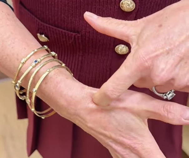
Where is Your Anatomical Snuffbox Located?

Anatomical Snuffbox location from Dr. Stacie Grossfeld

Key Takeaways:

  • The anatomical snuffbox is located at the base of the thumb, right on top of the scaphoid carpal bone.
  • Pain or tenderness at the anatomical snuffbox may indicate a scaphoid fracture.
  • Scaphoid fractures may require a splint, a cast, or surgery depending on the severity.

What is the Anatomical Snuffbox?

The anatomical snuffbox is the name of the triangular depression in the hand at the base of the thumb. It is bound laterally by the tendons of the abductor pollicis longus and extensor pollicis brevis muscles, and medially by the tendon of the extensor pollicis longus muscle. It sits right above the scaphoid, which is a carpal bone that connects to the distal radius, trapezium, and capitate.

Tenderness in the area of the snuffbox is often a sign of a scaphoid injury. The anatomical snuffbox is important because it hosts the radial artery, which is where a majority of the scaphoid’s blood supply comes from. If the scaphoid is injured, the amount of blood available to the injured scaphoid will determine how well it will heal on its own.

Scaphoid Fractures and Injuries

A scaphoid bone injury commonly results from falling on an outstretched hand. Symptoms may include:

  • Deep but dull pain at the wrist
  • Bruising of the wrist
  • Swelling of the wrist
  • Difficult gripping or squeezing

Because the pain may be dull, scaphoid injuries are often mistaken for a sprained wrist.

Treating Scaphoid Injuries

Treating scaphoid injuries will often depend on the severity and location of the injury. The blood supply to the scaphoid is inconsistent due to the shape of the bone, which makes healing inconsistent as well. Scaphoid fractures and other injuries are normally classified into three areas:

  1. Distal: the area nearest to the thumb
  2. Medial or Waist: the middle of the bone
  3. Proximal: the area nearest to the forearm

The distal pole gets the most blood supply and, which means that it has the best chance of healing without surgery. The waist and proximal pole do have a lot of access to the blood supply, so depending on the type of the fracture, it may require surgery.

Nondisplaced vs Displaced Scaphoid Fractures

The difference between nondisplaced and displaced scaphoid fractures is whether or not the break misaligns the bone pieces. If the bone has a gap or shifted pieces, it is unlikely the scaphoid will heal correctly without surgical resetting. Nondisplaced fractures will often heal well with a cast or splint.

Frequently Asked Questions About the Anatomical Snuffbox and Scaphoid

  1. What happens if a scaphoid fracture is left untreated?
    1. Scaphoid fractures that are not treated could lead to complications and possibly surgery. If the bone heals incorrectly and causes pain or deformation of the wrist, surgery will likely be necessary to fix it. Untreated scaphoid injuries can also lead to osteoarthritis of the wrist, carpal instability, and long-term pain.
  2. How are scaphoid injuries diagnosed?
    1. Like most fractures, imaging tests such as x-rays, CT scans, and MRIs may be used. However, because of the nature of the bone, multiple tests may be needed to identify a break. Some breaks may even be occult fractures, meaning that they do not show up well or at all on x-rays. Most doctors will use symptoms and snuffbox tenderness to rule scaphoid injuries in or out.
  3. How long does it take to recover from a scaphoid injury?
    1. Recovery time for scaphoid injuries averages 6-12 weeks with either a cast or surgery, but will vary with the severity and location of the injury. In some cases, rehabilitation may stretch to up to a year for a complete recovery.

If you or someone you love has suffered a scaphoid or anatomical snuffbox injury in the Louisville, Kentucky-area, board certified sports medicine physician Dr. Stacie Grossfeld at Orthopaedic Specialists PLLC can help. Orthopaedic Specialists PLLC is accepting new patients, and same day appointments are available. For additional information or to schedule an appointment, please contact Orthopaedic Specialists PLLC today at 502-212-2663.

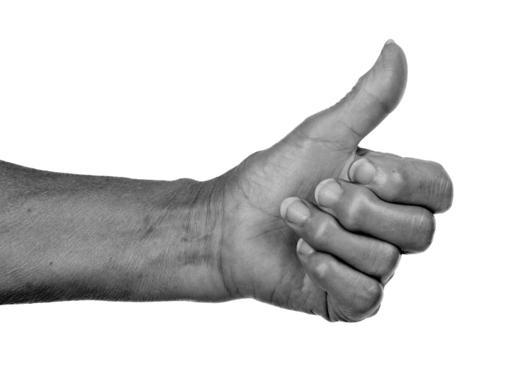
Hand Pain in Women: Carpometacarpal Arthritis

Carpometacarpal Arthritis of the Thumb

Key Takeaways:

  • The CMC (carpometacarpal) joint is at the base of the thumb, where the 1st metacarpal bone meets the trapezium.
  • The most common place women get arthritis in their hand is at the CMC joint.
  • Carpometacarpal arthritis is more common amongst post-menopausal women over the age of 50.

The carpometacarpal joint (CMC) is the basal joint of the thumb, right where the 1st metacarpal bone of the finger meets the trapezium bone of the hand. However, it is also the site of one of the most common reasons for hand pain in women over 50: carpometacarpal arthritis.

What is Carpometacarpal Arthritis?

Carpometacarpal arthritis is a type of osteoarthritis of the hand resulting from either trauma or wear-and-tear that decreases the amount of cartilage in the CMC joint. While this type of arthritis can be caused by trauma, it is most often the result of normal wear-and-tear as a joint that we use daily. The CMC joint is responsible not only for connecting the bones of the thumb to the hand, but also for its movement like swiveling, pivoting, grasping, and pinching.

With this type of repetitive movement, the cartilage that lubricates the joint and keeps the bones from grinding against each other eventually wears down. The pain, inflammation, swelling, and loss of motion that result are symptoms of carpometacarpal arthritis.

How are Women Affected by Carpometacarpal Arthritis?

Studies have shown that CMC arthritis is much more prevalent in women than men. According to Brown University Health, the disease affects 15% of adults over age 30, and two-thirds of women over the age of 55. The reasons for this can be because of genetics, lifestyle factors like weight, and environmental factors like how much and in what way you use your hand.

However, there may be biometrics, or measurable body characteristics, at play. Women have flatter joint surfaces, smaller areas of cartilage contact, and higher contact pressures. This leads scientists to believe that carpometacarpal arthritis may be more common in women due to a higher risk of altered joint loaded and decreased stability.

Women also have greater ligament flexibility. As we age and our ligaments become looser, our joints suffer from greater instability. After menopause, women then have a large hormonal shift. Studies show that the reduced estrogen levels affect collagen production, which affection the resiliency of joints.

How to Treat CMC Arthritis Pain

Carpometacarpal arthritis cannot be cured, but the pain can be managed like any other form of osteoarthritis. Common treatments include:

  1. Physical therapy: Strengthening the muscles of the hand and forearms can help stabilize the thumb joint and offload pressure when completing tasks.
  2. Thumb splints: When used correctly, thumb splints can help support the CMC joint during arthritic flare-ups.
  3. Corticosteroid injections: Injections are minimally-invasive and help to mitigate inflammation in joints affected by arthritis.

Normally, surgical intervention is not recommended unless there are special circumstances. Common surgical treatments for CMC arthritis may include joint fusions, joint replacements, or trapeziectomy (removal of the trapezium bone).

FAQs About Carpometacarpal Arthritis

  1. How does CMC arthritis affect daily activities?
    1. Those affected by osteoarthritis in the thumb may notice difficulty with tasks requiring grasping, pinching, or gripping. This might be opening bottles and jars, grasping doorknobs, playing sports like golf, tennis, or pickleball, and other activities.
  2. What are the symptoms of CMC arthritis?
    1. Symptoms of carpometacarpal arthritis can include pain at the CMC joint, inflammation, swelling, loss of grip strength, deformity of the joint, and loss of motion.
  3. Can arthritis affect metacarpal bones?
    1. While CMC arthritis pain can feel like the bones of your hand hurt, most pain will originate from the joint itself and the point where the bones are rubbing against each other. However, arthritis can affect any joint of the body, including those associated with the metacarpal bones.

If you or someone you love suffers from arthritis pain in the Louisville, Kentucky-area, board certified sports medicine physician Dr. Stacie Grossfeld at Orthopaedic Specialists PLLC can help. Orthopaedic Specialists PLLC is accepting new patients, and same day appointments are available. For additional information or to schedule an appointment, please contact Orthopaedic Specialists PLLC today at 502-212-2663.
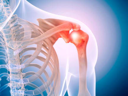
Shoulder Exam: How To Do a Crank Test for Labral Tears in the Shoulder

Shoulder Crank Test for pain

Key Takeaways:

  • The Crank Test is a non-invasive movement test that doctors can use to help identify labral tears in the shoulder.
  • This is only one of several movement tests, like the Mayo Shear test, that your doctor may perform to identify where pain is originating from.
  • Labral tears usually result from acute trauma or repetitive shoulder motion and are common in swinging or throwing athletes, or workers who lift heavy objects every day.

Similar to the Mayo Shear Test, the Crank Test is another movement-based test that requires no medical instruments and can be performed anywhere.

How To Do a Shoulder Crank Test

  1. Bring the patient’s arm to a 90 degree from their body
  2. Bend the elbow to 90 degrees, with the hand facing up
  3. Apply an axial load to the humerus (push the elbow into the shoulder)
  4. Internally and externally rotate the arm as you maintain the axial load

If you feel a clunk or a click, particularly when externally rotating the arm, there may be a labral tear in the shoulder.

What is the Difference Between a Crank Test and a Mayo Shear Test?

The three most common types of labral tears include:

  1. SLAP Lesion – a tear that occurs on the upper part of the socket. This kind of tear also affects the bicep tendon.
  2. Bankart Lesion – a tear that occurs at the bottom of the socket. This normally results from a shoulder dislocation.
  3. Posterior Labrum Tear – a tear that occurs at the back of the socket. Wear and tear or acute injuries are commonly the culprits for this type of tear.

Mayo Shear tests usually help to identify Bankart Lesions (Lennard Funk, Shoulder Examination Tests) and posterior labrum tears while Crank tests help to identify SLAP lesions. They utilize different movements and body mechanics, and the results target pain in different areas of the shoulder.

Frequently Asked Questions About the Crank Test and SLAP Tears

  1. What is a SLAP Lesion?
    1. A SLAP lesion is also known as a SLAP tear, which stands for Superior Labrum, from Anterior to Posterior tear. It occurs on the upper part of the glenoid socket. This kind of tear also affects the bicep tendon. Damaged or torn labrums often result from either acute trauma or repetitive shoulder motion (OrthoInfo). In many cases, this leads to shoulder pain (especially with overhead activities), shoulder instability, and other symptoms such as:
      1. Dislocations
      2. Sensations or noises of catching, locking, popping, or grinding
      3. Increased shoulder pain at night
      4. Decreased range of motion
      5. Loss of strength
  2. What are the next steps if a Shoulder Crank Test generates pain?
    1. If a Crank Test is causing pain, there is a possibility that you have torn your labrum. SLAP tears that are left untreated may lead to chronic shoulder pain and instability. If this test causes pain and a click, it is best to contact a doctor for a diagnosis. In the meantime, it is recommended that you rest your shoulder and abstain from activities that require overhead motion or heavy shoulder lifting.
  3. How is a SLAP lesion treated?
    1. SLAP lesions can take up to a year to heal, and can be treated either conservatively or surgically.
      1. Conservative treatments should be the first thing that a doctor prescribes. This can include rest or modified activity, physical therapy, and over-the-counter pain medication.
      2. Surgical treatments may be recommended if the pain gets to a point that it interferes with daily activities. The operation reattaches the torn labrum to the shoulder socket, which improves stability.

If you or someone you love has suffered a shoulder labrum tear in the Louisville, Kentucky-area, board certified sports medicine physician Dr. Stacie Grossfeld at Orthopaedic Specialists PLLC can help. Orthopaedic Specialists PLLC is accepting new patients, and same day appointments are available. For additional information or to schedule an appointment, please contact Orthopaedic Specialists PLLC today at 502-212-2663.
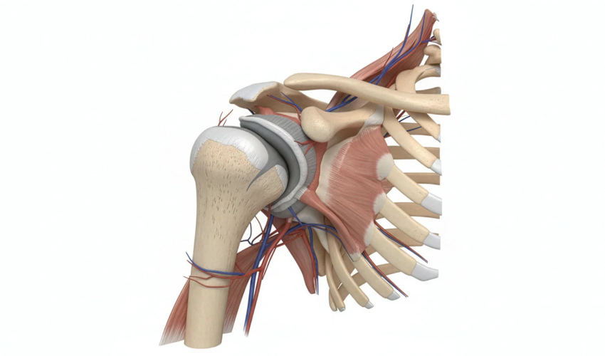
Shoulder Exam: How to Do a Mayo Shear Test for Labral Tears in the Shoulder

Shoulder Labrum Tears and the Mayo Shear Test

Key Takeaways:

  • A Mayo Shear Test is a noninvasive motion test that can help to identify labral tears in the shoulder.
  • If a labral tear is present in the shoulder, the patient will normally feel pain in the back of the shoulder and/or hear a clicking sound.
  • A labral tear occurs when the specialized cartilage tissue in the shoulder is torn or damaged, usually from throwing, pushing, or doing overhead tasks.

The labrum is a special piece of cartilage that cushions the ball-and-socket joint of the shoulder. This joint is made up of the humeral head from the arm (the ball) and the glenoid (the socket). The labrum lives on the rim of the glenoid and serves as an attachment location for the ligaments that connect the humerus and and the glenoid.

How Do You Know If You Have a Torn Labrum in Your Shoulder?

Damaged or torn labrums often result from either acute trauma or repetitive shoulder motion (OrthoInfo). In many cases, this leads to shoulder pain (especially with overhead activities), shoulder instability, and other symptoms such as:

  • Dislocations
  • Sensations or noises of catching, locking, popping, or grinding
  • Increased shoulder pain at night
  • Decreased range of motion
  • Loss of strength

There are three types of labral tears that are most common:

  1. SLAP Lesion – a tear that occurs on the upper part of the socket. This kind of tear also affects the bicep tendon.
  2. Bankart Lesion – a tear that occurs at the bottom of the socket. This normally results from a shoulder dislocation.
  3. Posterior Labrum Tear – a tear that occurs at the back of the socket. Wear and tear or acute injuries are commonly the culprits for this type of tear.

If you play a sport that involves a lot of throwing or a similar motion to it, or your daily activities require you to repetitively lift heavy objects, your shoulder pain might be a torn labrum. The only way to determine with any certainty that you have torn your labrum is with imaging tests like CT scans or MRIs. However, in order to narrow down the possible origin of your shoulder pain, doctors may perform simple, non-invasive movement tests like the Mayo Shear Test.

How To Do a Mayo Shear Test

For this labral tear of the shoulder test, you will:

  1. Adduct the affected arm
  2. Bend the elbow so the arm is at 90 degrees with the arm externally rotated and your hand on the back of their shoulder
  3. Bring up to about 120 degrees of forward flexion
  4. Then pull the arm down to about 60 degrees

If the up and down movement produces a click or other noise, and produces pain along the posterior (back portion) of the shoulder, they may have a labral tear, usually a Bankart lesion or posterior labrum tear.

Frequently Asked Questions About Labral Tears in the Shoulder

  1. Will a torn shoulder labrum heal completely?
    1. If the tear is mild and proper steps to rehabilitation is followed, most patients will have full function of the shoulder and return to their activities with few or no restrictions. In the case of Burkart lesions, some patients report feeling like their shoulder will dislocate again. Focusing on rebuilding the muscles of the shoulders will be very helpful for stabilization.
  2. Can a labral tear in the shoulder heal on its own?
    1. In some cases, conservative treatments like rest, over-the-counter pain killers, and physical therapy can be enough to heal a labrum tear. However, it is not recommended that no action be taken, as this will increase the possibility of reinjury, or worsening the tear.
  3. How do you fix a labral tear in the shoulder?
    1. Common treatments for shoulder labrum tears include activity restrictions, physical therapy, anti-inflammatory medication, corticosteroid injections for pain management, or in more severe cases, shoulder arthroscopy surgery to trim or reattach the labrum to the glenoid.
  4. How long does it take to recover from a labrum tear in the shoulder?
    1. Without surgery, this will vary case-by-case. Healing can be unpredictable due to factors like how rigidly the patient adheres to a recovery plan and how severe the tear is. With surgery, there is more structure, and patients usually have a minimum recovery period of 12 weeks, but it will take 4-6 months to return to activities at full-strength.

If you or someone you love has suffered a shoulder labrum tear in the Louisville, Kentucky-area, board certified sports medicine physician Dr. Stacie Grossfeld at Orthopaedic Specialists PLLC can help. Orthopaedic Specialists PLLC is accepting new patients, and same day appointments are available. For additional information or to schedule an appointment, please contact Orthopaedic Specialists PLLC today at 502-212-2663.

Shoulder Exam: How To Do a Shoulder Adduction Test for Acromioclavicular Pain

Shoulder Adduction Tests and AC Joint Pain Shoulder Diagram

Key Takeaways:

  • The shoulder is made of two joints: the glenohumeral joint and the acromioclavicular joint.
  • The acromioclavicular joint is where your collarbone connects to your shoulder blade.
  • A shoulder adductor test can help identify if shoulder pain originates from the acromioclavicular joint.

If you are feeling pain in your shoulder, your doctor may conduct a shoulder adductor test. Because the shoulder is made up of two joints, the glenohumeral joint and the acromioclavicular joint, this test can help identify which of them may be producing the pain. A shoulder adduction test determines if someone has acromioclavicular pain, or pain at the AC joint.

The shoulder adduction test is very simple:

  1. The patient will raise their hand and arm 90 degrees in front of themselves.
  2. A doctor will move it across the chest horizontally, or adduct it.
  3. If there is pain can be reproduced in the AC joint area, the test is positive.

What is the Acromioclavicular Joint?

The acromioclavicular joint, also known as the AC joint, is one of two joints in the shoulder. It connects the clavicle (collarbone) to the top part of the scapula, called the acromion (Washington University Medicine). When you move your shoulder, the AC joint helps it move from back to front as a planar joint, or a joint that lets two bones glide past each other.

Its primary job is to support your shoulder and act as a connection point for the surrounding ligaments. It is a passive joint, so it does not move by itself. Any movement of the AC joint is performed by the muscles, tendons, ligaments, and the other joints. The AC joint includes the following components:

  • Collarbone
  • Shoulder blade
  • Cartilage
  • Nerves
  • Blood vessels
  • Acromioclavicular ligament
  • Coracoclavicular ligament, made up of the trapezoid and conoid ligaments

Most Common Ways the AC Joint is Injured

The AC joint is susceptible to injury from both the shoulder and the collarbone. Stretched, strained, or torn ligaments are possible, especially with acute shoulder trauma associated with (Mass General Brigham):

  • Falling directly on the shoulder
  • Sustaining a hard hit to the shoulder
  • Lifting heavy objects

Arthritis can also develop if the cartilage sustains damage or wear-and-tear. The most common sport injuries for AC joints are heavy-hitting sports like American football, martial arts, hockey, skiing, snowboarding, and biking.

Degrees of Injury to the AC Joint

There are 6 classifications for injury to the AC joint and its ligaments (National Library of Medicine, Acromioclavicular joint injuries: diagnosis, classification and ligamentoplasty procedures):

  • Type I is a sprain injury of the AC ligament. There is no complete tear and both AC and CC ligaments are intact.
  • Type II is a tear of the AC ligament but not of the CC ligaments.
  • Type III involves tears of both the AC and CC ligaments, with 25% to 100% displacement of the clavicle compared with that on the contralateral side.
  • Type IV is when both the AC and CC ligaments are torn and there is posterior displacement of the distal clavicle into the trapezius fascia.
  • Type V involves the AC and CC ligaments and both the origin of the deltoid and insertion of the trapezius tearing, causing extreme instability of the AC joint. It is a complex injury where the deltotrapezial fascia is stripped from its attachment and displacement of the clavicle is more than three times the diameter of its distal part. The CC distance is increased to 100% to 300%.
  • Type VI injuries are the result of inferior displacement of the distal clavicle into the subcoracoid position.

Frequently Asked Questions About the AC Joint and Shoulder Adduction Tests:

  1. How accurate is the shoulder adduction test?
    1. A shoulder adduction test is a non-invasive tool that people can use to help get a sense of where pain is coming from. It is not meant to formally diagnose any specific condition, so accuracy depends mostly on pinpointing the origin point of pain and then taking the correct steps.
  2. What should you do if a shoulder adduction test causes pain?
    1. If you perform a shoulder adduction test and find that you have pain at or around the AC joint, the next steps would be to identify how much pain you are in and make a decision to see a doctor or treat the symptoms yourself. However, if treating your AC joint pain with OTC pain medications, ice, and rest does not help within a few days, see a doctor.
  3. What AC joint conditions does a positive shoulder adduction test implicate?
    1. A positive shoulder adduction test does not implicate a specific condition. However, common injuries and conditions that may cause your test to be positive include arthritis, sprains, torn ligaments, or a dislocated joint.

If you or someone you love has suffered an AC joint injury in the Louisville, Kentucky-area, board certified sports medicine physician Dr. Stacie Grossfeld at Orthopaedic Specialists PLLC can help. Orthopaedic Specialists PLLC is accepting new patients, and same day appointments are available. For additional information or to schedule an appointment, please contact Orthopaedic Specialists PLLC today at 502-212-2663.