Under Pressure: What Causes a Pinched Nerve?

Nerves act as sounding alarms for our bodies, sending messages to alert us of damage done. In the case of a pinched nerve, we often feel sharp pain. This pain is the symptom of nerve compression.

What is a Pinched Nerve?

When a nerve is surrounded by too much compression from nearby tissues such as muscles, tendons, cartilage or bones, it becomes pinched from the pressure. Even the simplest of actions can cause a pinched nerve; whether it be lifting something too heavy or bending at the wrong angle. 

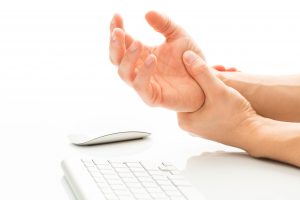
It’s normal if this pain is felt at the site of the compression as well as throughout your body as our nerve paths exist in our back, neck, arms and legs. In addition to pain, one may experience numbness, weakness and a tingling sensation as a result of a pinched nerve. Carpal tunnel syndrome for example, can be caused by a pinched nerve in the wrist; leading to pain and numbness in your fingers and hand.

Causes of a Pinched Nerve

An injury such as a herniated disk can also cause a pinched nerve to occur by putting excessive pressure on the root of the nerve. Being overweight is another condition that can result in a compressed nerve, as well as bone spurs and repetitive physical movements either from sporting activities or labor intensive tasks. Additionally, inflammation from arthritis can even cause tissue to compress nerves in our joints. 

Treating a Pinched Nerve

While a pinched nerve is usually only felt for a short period of time, it can lead to chronic pain and permanent nerve damage if left untreated. Treatment usually includes rest from the activity thought to have caused the tension in the first place as well as physical therapy to relieve the tightness, and stretch and strengthen your muscles for greater flexibility. Steroid injections can also be utilized to reduce any inflammation near the nerve.

Habits like practicing good posture, not staying in the same position for too long, and taking breaks from repetitive physical activity can reduce your chances of experiencing a pinched nerve and over stressing a specific area of your body. 

Surgery may be required if the pressure on the nerve is not relieved. If you or someone you know is suffering from a pinched nerve, one should seek treatment as soon as possible in order to alleviate any pain and prevent the nerve damage from worsening.

If your pinched nerve symptoms persist, contact Dr. Stacie Grossfeld.  With over 25 years of experience in orthopedic medicine, Dr. Grossfeld has extensive knowledge surrounding musculature. If you are suffering from prolonged pinched nerve symptoms, contact Orthopaedic Specialists today at 502-212-2663.

Common Sports Injuries For Kids: Learn More from Louisville Children’s Orthopedic Surgeon Stacie Grossfeld


It’s important as parents, coaches and mentors that we encourage kids to live an active healthy life. This naturally includes participation in sports. However, all physical activities come with the risk of injury, especially as kids are prone to tumbles, scrapes, bumps and bruises.

Being prepared for potential sports injuries is critical in that fast and effective treatment can often limit the severity of the injury. That being said, here are 5 of the most common sports injuries for kids and preventative measures you can take to avoid them.

1. Heat Related Illness

Kids can experience dehydration and or heat exhaustion if they are over exerting themselves in particularly hot weather coupled with high humidity. Factors such as body weight and not being accustomed to physical activity in the heat can increase the chances of a heat related illness. Signs include headache, stomach pain, nausea and thirst. All children participating in sports should practice proper hydration to prevent such an event from occurring, 

2. Shoulder Dislocations

Shoulder dislocations happen when the arm is forced backwards and one hears a pop. The result is immense pain and limited movement. To ensure that the shoulder heals properly, only a trained physician should put it back in place. Young athletes tend to be prone to these injuries as their joints are looser. However, physical therapy can help strengthen shoulder muscles and prevent a repeat dislocation.

3. Torn ACL

A brunt force to the outside of the knee or sudden and extreme movement from the athlete themselves can cause a torn ACL. One will often feel a popping or tearing feeling in the knee and have trouble bearing weight. As most ACL tears require surgery, one should consult with an orthopedic surgeon or sports medicine physician as soon as possible.

4. Concussion

There are over 250,000 sports related concussions per year among student athletes. They are most common in contact sports. Symptoms include headaches, slurred speech, mood swings, loss of memory, nausea and vomiting, dizziness, as well as confusion. While concussions are a serious brain injury, they can easily be left undetected. Proper concussion protocol must be followed after an event with head trauma occurs. The only treatment for a concussion is rest from physical activities and electronic devices for a long period of time before returning to play. 

5. Sprains & Strains

Injuries to the hand, wrist and ankle are all too common in active children. Most are the result of a fall. Swelling, pain and lack of movement are sure signs of an injury. However, immediate rest, icing, compression and elevation tend to be most effective for both sprains and strains of any kind. Sprains affect ligaments, while strains impact muscles and tendons. To determine if it is a break or fracture, an X-ray must be preformed and a doctor will decide if a cast or bracing is needed.

It should be noted that over use injuries in one area of the body can be avoided by mixing up the sporting activities a child participates in. Additionally, kids are at more of a risk for injury if they aren’t used to being active as their body is not conditioned for physical activity. 

Dr. Grossfeld has over 25 years of experience in orthopedic medicine and is a double board-certified orthopedic surgeon in sports medicine. To get more information about sports related injuries, contact Dr. Stacie Grossfeld today by calling 502-212-2663 to make an appointment. Orthopaedic Specialists has two convenient locations in Louisville, KY and is currently accepting new patients.

Important Facts About Sports Related Concussions in Female Athletes

There are over 250,000 sports related concussions per year among student athletes. However, the female athlete has been shown to sustain 50% more concussions than the male athlete. The differences are most commonly seen in sports such as basketball, soccer, and volleyball, as well as lacrosse.

Additionally, some studies have documented that female athletes have more severe concussions then their male counterparts. This is often coupled with worse symptoms, including greater cognitive functional deficits as well as a longer recovery period.

Why do females have a higher risk of sustaining a concussion? There have been many medical theories proposed to answer that question…

  1. Females have slender necks and smaller heads compared to males. Biomechanical studies demonstrate that females have 50% more head exhilaration during head trauma than males. This  translates into a higher likelihood of injury to the brain during a traumatic event.
  2. The female athlete has a relatively weak neck musculature compared to males. This may lead to less protection of the head during a major impact.
  3. Hormonal differences could also play a role as estrogen has been shown to be more detrimental to a brain after an injury.
  4. Additionally, there is a theory that the male athlete tends to report concussions less than a female does. There have been reports that gender stereotypes reinforce this behavior as males may try to provide a tougher persona. However, in my experience taking care of the University of Louisville women’s soccer and lacrosse team, I would disagree with this theory.

Dr. Grossfeld has over 25 years of experience in orthopedic medicine and is a double board-certified orthopedic surgeon in sports medicine. To get more information about concussions and other sports related injuries, contact Dr. Stacie Grossfeld today by calling 502-212-2663 to make an appointment. Orthopaedic Specialists has two convenient locations in Louisville, KY and is currently accepting new patients.

The Top 7 Things You Can Do To Avoid Knee Replacement Surgery

Knee problems are becoming more and more prevalent for patients of all ages, as old sports injuries rear their ugly head. However, even for those in constant pain, going under  the knife should never be the first option. There are many treatment options that can add longevity to your knees and have been found to be just as effective as surgery.

7 Ways You Can Avoid Knee Replacement Surgery 

  1. Weight loss. For every pound less that you weigh, you will place 3 pounds less of force on your knee joint when you walk.
  2. Physical therapy. When you strengthen the muscles around an arthritic joint, it can help to reduce the force and pain on the joint. There are some exercises that are very hard on an arthritic knee. A physical therapist is a good resource when seeking guidance for the correct exercises.
  3. Anti-inflammatory medicine. An oral anti-inflammatory medication such as ibuprofen or Aleve can be helpful. However, a primary care physician should determine if you are a good candidate before hand. As anybody with a history of renal disease or stomach ulcers cannot take this class of medication.
  4. Unloader knee brace. 90% of people‘s new arthritis is located in the inner part of their knee (also known as the medial compartment). An unloader knee brace can force you to place more weight on the outer half of your knee which typically does not have the same degree of arthritis. This can help to significantly reduce pain from arthritis and theoretically lessen the progression of the arthritis or at least slow it down.
  5. Cortisone injection. Cortisone is a steroid that is known to help reduce inflammation in the inner part of the knee joint. However, it is only temporary and tends to work between 3 to 6 months
  6. Hyaluronic injection. Hyaluronic acid is a natural fluid in your knee joint. When you get arthritis, this fluid becomes very thin and does not provide a cushion to the knee. Injecting a synthetic hyaluronic acid tricks the knee joint into producing a more normal fluid for about 6 to 12 months. This can help reduce knee pain for that duration of time in an arthritic knee.
  7. PRP injection. PRP stands for platelet rich plasma. Platelets have a significant amount of growth factors. Blood is drawn from your arm, centrifuged, and then platelets are injected back into the knee joint. This can give you up to 12–24 months of pain relief in an arthritic knee.

If you are suffering from joint pain due to inflammation, arthritis, or an injury — contact Dr. Stacie Grossfeld today by calling 502-212-2663. Dr. Stacie Grossfeld is an orthopedic surgeon in Louisville, KY who is double board-certified in orthopedics and sports medicine. She has over 20 years of experience in orthopedics and is always looking for new treatment methods to bring to her clientele. Orthopaedic Specialists has two convenient locations in Louisville, KY and is currently accepting new patients.

Are You a Candidate for Subchondroplasty?

When your bones experience continuous wear and tear, they can develop stress fractures or bone marrow edema (also referred to as bone bruises or chronic bone marrow lesions). This is quite common in older adults; specifically women and can cause pain as well as physical limitations.

A new joint preserving procedure called Subchondroplasty, provided by a board certified orthopedic surgeon like Dr. Stacie Grossfeld, may be able to relieve a patient’s pain and restore joint function. Subchondroplasty entails an injection of a hard-setting bone substitute that mimics the chemical structure of an actual human bone. Once injected into the bone, it works to seal up a fracture and over time can even be resorbed and replaced with new bone. Subchondroplasty is a minimally invasive treatment and oftentimes spurs bone growth resulting in a healed bone and reduced pain in the leg, knee or hip.

Are you a Candidate for Subchondroplasty?

If you have suffered from chronic and severe bone pain for at least 3 months and have trouble walking or standing, you could benefit from Subchondroplasty. If other injections, anti-inflammatory or pain medications, knee braces, crutches, physical therapy, and or rest have not worked, Subchondroplasty could be the right treatment. Additionally, patients with cartilage loss and bone deterioration associated with osteoarthritis, as well as those diagnosed with arthritis or osteoporosis are usually good candidates for the procedure.

An MRI is first required to determine if you have bone marrow edema, as spots can be seen in the bone that represent swelling or bone damage. A relatively healthy body weight and or lifestyle changes are suggested for potential patients receiving this treatment in order to prevent reoccurring problems that could require further treatments.

Subchondroplasty is a popular choice for patients who enjoy living an active life and want to avoid a bone replacement. It is an outpatient procedure, usually lasting 20 minutes. While it depends on the patient, most can bear weight immediately after treatment is complete. 

If you or someone you know is experiencing pain due to insufficiency fractures or bone marrow edema, contact Dr. Stacie Grossfeld at Orthopaedic Specialists. Dr. Grossfeld has been performing subchondroplasty for about two years and has had excellent results. For more information and to schedule an appointment with Louisville orthopedic Dr. Stacie Grossfeld, call 502-212-2663. You can also visit either of the Orthopaedic Specialists’ locations in Louisville, KY to get more information and schedule an appointment today.

Pitting Edema VS Non-Pitting Edema: What’s the Difference?

pitting edemaEdema is a medical terms that refers to swelling caused by the body holding onto fluid. Edema, which was once called dropsy, usually occurs in a person’s feet, ankles, or legs. There are some occasions where edema can also occur in a person’s hands, face, or even affect additional body parts. There are many different kinds of edema and the causes can vary from diagnosis to diagnosis. Edema is often a symptom of another, more serious, condition and should be treated by a medical doctor. Two common forms of edema are pitting edema and non-pitting edema. These are the most common classifications of edema and are easily identifiable.

The biggest difference between pitting and non-pitting edema is the way the two conditions respond to pressure. Pitting edema responds to pressure, be it from a finger or a hand, while pitting edema does not. If you press on your skin with your finger and it leaves an indentation, you could be suffering from pitting edema. Non-pitting edema, on the other hand, does not respond to pressure or cause any sort of indentation. Follow along to learn more about pitting and non-pitting edema including causes, treatments, and more information.

Pitting Edema

Pitting edema often affects areas of the lower body including the feet, legs, and ankles, but can occur anywhere on the body. This condition is often a local problem that involves the veins, but can also be a problem within one of your body’s systems of organs. Pitting edema can be a result of problems with the liver, kidneys, heart, or even the lymphatic system. Regardless of where the pitting edema is occurring, it is a sign that your body is holding onto fluid or fluid is not being carried away as it should. If these symptoms apply to you, you should seek medical help immediately.

While pitting edema can usually be associated to a problem with the liver, kidneys, heart, or lymphatic system, there are many other causes that can result in pitting edema. Some of those various issues include:

  • Lung Diseases
  • Obesity
  • Pregnancy
  • Medications
  • Low Levels of Protein
  • Heart Failure

Some of these medical conditions are not cause for concern. Pitting edema in pregnancy is more common than not; however, it can still be a cause for concern and should be mentioned to your doctor. Certain medications that result in pitting edema should be reassessed by the prescribing doctor. Risk factors that are associated with pitting edema include: high sodium intake, emphysema, immobility, sedentary lifestyle, and more.

Diagnosis for pitting edema is somewhat difficult since a list of telltale signs for diagnosing this medical issue does not exist. However, through a set of tests and an extensive evaluation that may include a chest x-ray, blood work, and a heart echo can help a physician identify pitting edema. If you are diagnosed with pitting edema, there are a wide variety of treatments available. Some treatments can be applied at home while others must be performed by a practicing physician.

Non-Pitting Edema

Non-pitting edema is similar to pitting edema save for the indentation (or “pit”) that remains visible after pressure is applied in a case of pitting edema. Unlike pitting edema, non-pitting edema is much easier to diagnose because it is caused by three very specific conditions: lymphedema, myxedema, and lipedema.

Lymphedema

When it is extremely difficult for lymph fluid to drain as it should, it is often due to a blockage. This condition is called lymphedema. Some people are born with this condition while others develop it. Those who develop lymphedema often develop it after surgery or as a secondary condition to a bigger issue. When this lymph fluid cannot drain, it gathers and swelling results.

Myxedema

Another condition that can be linked to pitting edema is myxedema. Myxedema can be identified by severe swelling in the legs and feet, but swollen eyelids and lips are also symptoms. Tongue swelling is another symptom of myxedema though it is not as common as the swelling observed in other areas of the body. Most people who suffer from myxedema suffer from hypothyroidism – both severe and advanced cases.

Lipedema

Lipedema is another condition that results in non-pitting edema. When fat cells grow at an increased rate, they tend to retain more fluid. This results in a painful case of non-pitting edema that occurs in women, almost exclusively.

These three causes of non-pitting edema can be diagnosed by unusual swelling. Once a doctor identifies unusual swelling, they will review medical records and do a wide variety of tests to diagnose. Among the three medical conditions, lipedema is the hardest to diagnose and requires a physical examination. During the exam, doctors will look for: tender skin, excessive weight, and bruising. Once non-pitting edema is diagnosed, depending on the condition associated with it, the treatment plans vary and are determined by a trained physician.

Dr. Stacie Grossfeld is a trained orthopedic surgeon who is double board-certified in orthopedic surgery and sports medicine. If you feel that you are suffering from abnormal swelling, or have symptoms that align with pitting edema, reach out to Dr. Stacie Grossfeld today. You can contact her by using the contact form on her website or by calling 502-212-2663 today!

5 Treatments for Golfer’s Elbow

If you have been experiencing pain when gripping or arehaving weak wrist movement in general, it’s possible you are experiencing what is called golfer’s elbow. Golfer’s elbow is known medically as medial epicondylitis and can sometimes even be a form of chronic tendinitis. Golfer’s elbow is also commonly referred to as “thrower’s elbow.” It is caused by repeated forceful motions like throwing, lifting, or bending the wrist against a golf club improperly. Golfer’s elbow affects more than just golfers. Those of all occupations or recreational hobbies can be susceptible if they’re bending their elbow often, and not weight training correctly or taking the time to warm up.  

Golfer’s elbow is easily confused with tennis elbow and is similar in that it is caused primarily from overuse. However, golfer’s elbow affects the inside of the elbow rather than the outside. Pain occurs at the point where the tendons of your forearm muscles meet the inner bone of your elbow. This results in “damage to the muscles and tendons that control your wrist and fingers.”

Golfer’s elbow can easily be prevented. If you are an avid golfer, golfer’s elbow surprisingly affects the trail arm of the swing more than the lead arm. It is often developed from golfers who tend to release their wrists too soon during their down swing. To help correct this issue, consider taking lessons to correct your form and avoid playing on poorly maintained courses that have too hard of grounds. A slower swing may even help reduce the amount of blunt force to the joint when hitting the ball.

Rest is key in treating golfer’s elbow.

It is imperative that you take a time out from any movement that may have caused the injury in the first place. Not refraining from physical activity will most likely prolong the healing process. Once you are healed, make sure to make a gradual re-entry to your normal activities.  

Other self treatment methods include:

  • Propping the elbow up in order to keep it elevated. This helps stimulate blood flow.
  • Icing the area as much as four times a day for 20 minutes can also be beneficial. However, if this is a recurring problem heat therapy might be more conducive.
  • Over the counter anti-inflammatory medicine can aid in relieving both pain and reducing inflammation. However, consulting with your healthcare provider is necessary before taking any medication. To reach a Kentucky orthopedic, feel free to contact Dr. Grossfeld at Orthopaedic Specialists at 502-212-2663

5 Treatments for Golfer’s Elbow

  1. Utilize a splint or brace. These can strengthen as well as protect the tendon and muscles affected; removing some of the strain and tension.
  2. Rehabilitation exercises such as squeezing a tennis ball, or doing wrist curls (curling a small weight from your fingers to your arms) can be effective at strengthening the forearm muscles.
  3. A deep tissue massage as part of an occupational therapy treatment plan can result in immense pain relief.
  4. Cortisone injections offer pain relief and reduce inflammation as well. However, they are not an effective long-term treatment option as many return too quickly to the activity that caused injury in the first place.
  5. Though platelet rich plasma (PRP) injection therapy is a relatively new concept, it has been shown to treat chronic tendon injuries quickly. PRP injections are non invasive. They involve drawing a small amount of blood from the patient, running it through the process of centrifugation, and then injecting it into the inflamed tissue. Dr. Stacie Grossfeld currently offers PRP injection therapy. Call 502-212-2663 today for more information or to schedule an appointment.

If not treated, golfer’s elbow can limit your range of motion and result in chronic pain.

While treatment for such an injury ranges from patient to patient, the recovery period is often a few weeks, with more severe cases taking months to heal. Dr. Stacie Grossfeld, an orthopedic surgeon and elbow specialist, in Louisville, Kentucky recommends seeking medical treatment as soon as the pain starts in order to avoid further damage and or chronic pain. In the rare case that you’ve torn the tendon you may need surgery to repair it.

If you are suffering from elbow pain or have recently injured one of your elbows, contact Dr. Stacie Grossfeld immediately. Dr. Grossfeld is a double board-certified orthopedic surgeon with a focus in sports medicine who routinely treats and repairs elbow injuries. Dr. Grossfeld can be reached at either of her two office locations in Louisville, Kentucky by calling 502-212-2663.


5 Yoga Poses That Help Frozen Shoulder Recovery

frozen shoulderYoga originated in ancient India and includes a wide variety of schools and practices. The goals of these schools and practices vary, as well. With the recent rise in interest in yoga in the Western world, more goals and benefits of yoga are being discovered including health benefits and rehabilitation benefits. Recently, it was discovered that some yoga poses even help orthopedic issues including frozen shoulder.

What is a Frozen Shoulder?

Adhesive capsulitis, or “frozen shoulder,” is characterized by pain and stiffness in the shoulder. It affects about 2 percent of the population; most commonly it is found in women and people ages 40 to 60. Frozen shoulder may be caused by a health problem that causes general immobilization. The shoulder may also freeze as a way to protect itself and prevent further injury after a trauma.

There are three phases of frozen shoulder: freezing, frozen, and thawing.

  1. Freezing – Typically lasting for six weeks to nine months, this phase is characterized by a gradual onset of pain in the shoulder. As the pain becomes more intense, the shoulder loses its full range of motion.
  2. Frozen – This phase occurs in the four to six months after the freezing. Your pain will decrease, but your range of motion will still be limited. Daily activities become difficult.
  3. Thawing – As the shoulder slowly regains its range of motion, it is in the thawing stage. It takes somewhere between six months and two years for the shoulder to regain its normal– or close to normal– strength and range of motion.

Yoga Poses for Frozen Shoulder

Yoga is an exercise practice that combines breath control, simple meditation, and poses to help relax and strengthen the mind and body. When practicing yoga, always listen to your body and breath. If you push yourself to a point where you lose concentration and stop breathing, you are being too aggressive to your injury.

Below is a yoga routine utilizing five simple poses that can help your frozen shoulder recover and reach the thawing phase.

1. Start on your hands and knees with your back straight. Take a deep breath in and lift one arm straight in front, keeping your back from curving or caving. Exhale and return to your hands and knees. Repeat on the other side.

2. From your hand and knees, tuck your toes in and raise you hips to downward facing dog. Take a few large breaths, in and out, in this position.

3. Release your knees and come down on your stomach. Gently and slowly, reach your hands to your ankles or toes for bow pose. Take a few large breaths, in and out, in this position.

4. Slowly lower your arms and legs back to the ground then rise to sitting on your knees with your hands in a prayer position and take a few breaths. Breathe in as you place one foot in front of your body and turn into half-prayer twist pose. Exhale to release tension in that shoulder. Return to knees and prayer pose then repeat on the other side.

5. With both sides stretched, return to knees on ground. Breathe in and raise arms overhead. Exhale slowly as you circle your arms down and around and place your hands behind you in reverse prayer position. Take a few big breaths, in and out, in this position. Once done, you can move to child’s pose to finish the practice.

Frozen shoulder is a serious orthopedic issue.

However, this yoga flow will help with recovery and thawing your frozen shoulder. Speak to your doctor today about how you can incorporate yoga into your rehabilitation routine.

If your frozen shoulder symptoms persist, contact Dr. Stacie Grossfeld.  With over 10 years of experience as an orthopedic surgeon, Dr. Grossfeld has extensive knowledge surrounding shoulder injuries and rehabilitation. If you are in the Louisville or Kentuckiana area and are suffering from frozen shoulder, contact Orthopaedic Specialists today at 502-212-2663.

10 Facts about Rotator Cuff Injuries and Repair

Your shoulders are important body parts that should be treated with care; however, the amount of work you make your shoulder do every day often goes unconsidered. This can sometimes result in an injury. Rotator cuffs are involved in most shoulder injuries. Rotator cuff injuries and tears are one of the most common injuries to effect adults and athletes.

rotator cuff injuriesThe rotator cuff is “a group of four muscles that come together as tendons”. These tendons “form a ‘cuff’ over the head of the humerus”. The four muscles that create this cuff are the supraspinatus, infraspinatus, subscapularis, and teres minor. These muscles all originate from the scapula, collar bone, and attach to the humerus. They attach at specific spots known as the greater tuberosity and lower tuberosity.

The main functionality of the rotator cuff is to lift and rotate the arm. It also stabilizes the ball of the shoulder within the ball and socket joint that allows free movement of the arm.   However, the rotator cuff has other uses and rotator cuff injuries affect those significantly. Below are ten facts surrounding rotator cuffs and rotator cuff injuries including the most common – rotator cuff tears.

Rotator Cuff Injuries, Tears, and Repairs: 10 Important Facts

  1. Tears are found in 30% – 50% of the population that is older than 50.
  2. Rotator cuff tears are among the most common shoulder disorders that require surgical management.
  3. Once the rotator cuff tendon tears off of the humerus bone, it starts to lose its nutrition. This causes it to begin to die. Timely surgery can greatly improve rates of surgical repair.
  4. Risks for post-operative failure after rotator cuff surgery include: a tear that is larger than 2.5 cm, fatty degeneration of the muscles in an MRI scan, older patient age, patients who are smokers or diabetics with blood sugar control.
  5. Fatty degeneration is associated with muscle weakness after shoulder surgery. This affects shoulder function in addition to being a risk factor for post-operative failure.
  6. After rotator cuff repair surgery, tendon retreat rates of anywhere between 25% and 95% have been reported by orthopedic surgeons observing post-operative patients.
  7. PRP (platelet rich plasma) has been shown to enhance the quality of early tendon healing in rotator cuff repair surgeries.
  8. The supraspinatus is the most common muscle torn out of the four muscles that make up the tendons in the rotator cuff.
  9. Once the rotator cuff surgery has been performed and rehabilitation is completed, a patient will have spent 5-6 months towards total recovery.
  10. Rotator cuff surgery will restore the entire range of motion to the shoulder. It will also reduce pain and significantly improve function when successful.

Injury to a rotator cuff can occur due to sports, sustaining a fall, or can simply be age-related.

Most commonly, a rotator cuff repair is diagnosed with an MRI after a patient complains of severe pain in the shoulder area and visits an orthopedic surgeon. Once an MRI is completed and the pain is diagnosed as a rotator cuff injury then treatment is decided. Not all rotator cuff repairs require surgery; however a large rotator cuff tear often requires surgery and months of rehabilitation in order to properly heal and regain functionality.

One of the most important things you can do if you suffer a rotator cuff injury is to seek medical attention immediately. Recovery rates are drastically improved when a rotator cuff injury is diagnosed and treated by a doctor as quickly as possible. If you feel that you may have an injured rotator cuff, contact Dr. Stacie Grossfeld at Orthopaedic Specialists immediately by calling 502-212-2663.

Injection Therapies for Knee Osteoarthritis

Platelet Rich Plasma (PRP)

PRP injections are a process where blood is drawn out of your arm in the office. We then centrifuge your blood in order to separate out the platelets from the rest of your blood cells. These platelets are then injected back into the knee joint. PRP injections tend to work as well or better than a hyaluronic acid injection. The entire process takes about 30 minutes and requires a blood draw of approximately 10 cc.

Hyaluronic Acid (HA)

Hyaluronic Acid is a synthetic fluid that is injected into the knee and helps to stimulate the synovial cells that line the inner part of the knee joint. This helps produce a more thick cushion-like fluid for the knee. In a normal knee, the fluid in the knee joint is thick like motor oil. However, when the knee becomes arthritic, it starts producing a fluid that has a water like consistency. After injecting the hyaluronic acid into the knee, it “tricks” the knee joint into producing a thicker more viscous joint fluid. Effects of HA injections usually last up to 6-12 months. Typically most insurance companies will cover a percentage of the cost.

Cortisone

Cortisone is a steroid that helps reduce inflammation temporary and is excellent for acute pain relief. It lasts between 3 to 4 months and does not have any growth factors. However, during the 3-4 month time frame, the cortisone will wear off and the symptoms will return. Cortisone shots are usually covered by your insurance company.

When it comes to treating an arthritic knee joint and chronic pain, there are several options we can provide that range in both efficacy and price. Prior to receiving any treatment, your coverage will be checked as benefits with insurances companies tend to change on a regular basis. To get more information on what would be the best treatment option for you, make an appointment with Dr. Stacie Grossfeld at Orthopaedic Specialists in Louisville, KY by calling 502-212-2663. Dr. Grossfeld has over 25 years of experience in orthopedic medicine and is a double board-certified orthopedic surgeon.