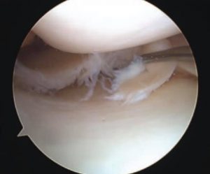
A meniscus is a structure located in your knee joint that functions as a shock absorber. You have two of them in each knee and they are prone to tearing. When the meniscus is torn, it produces a sharp stabbing pain and sometimes it will cause your knee to click or even lock. Many times swelling is associated with a meniscal tear.
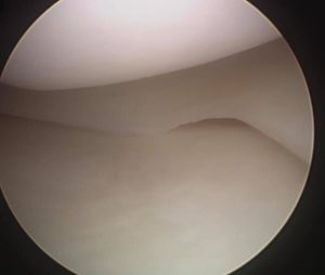
The treatment for a meniscal tear is a knee arthroscopy. That is the procedure where I go in and remove the torn part of the meniscus. On occasion the meniscal tear can actually be repaired or sutured back together but that is only rare. The reason why the meniscal tears are typically not repaired is because the meniscus does not have a good blood supply. So if I place sutures in the meniscus it would just not heal. There is a part of the meniscus that is very vascular and has a good blood supply. If the tear occurs in that area then the meniscus can be repaired versus just removing the part that is torn.
The meniscus has shock absorbing qualities but once it is torn, the area where the tear is located no longer provides any shock absorbency. The only thing the torn meniscus provides is pain.
Often when a patient comes in with back pain, they ask the question, “Do I have scoliosis?”
Scoliosis is a curvature and a rotational deformity of the spine. By definition, the curvature of the spine should be at least 10°.
There are 2 Types of Scoliosis:
1. Adolescent Onset Scoliosis
The standard case of scoliosis usually occurs during a growth spurt right before puberty. However, it is possible that the condition may not be caught until adulthood when the symptoms become more problematic. While the cause of adolescent onset scoliosis can be attributed to birth defects, a spinal injury, muscular dystrophy, or cerebral palsy, the cause of most pediatric cases is unknown. It has also been determined that scoliosis can be genetically related.
Treatment is based on the age of onset and the degree of the curve. This can range from simple observation and bracing, to complex invasive surgery. If caught early on, the issue can most likely be resolved. Uneven shoulders, hips or waist are signs of scoliosis.
2. Adult Onset Scoliosis
Also known as degenerative scoliosis, this form of the condition typically affects those 50 or older. Adult onset scoliosis can slowly occur over time with age. In fact, it is normal to experience a form of degeneration when it comes to facet joints and intervertebral discs. As we age, this kind of deterioration is also known to cause osteoarthritis as well as degenerative disc disease.
According to a recent study, “at least 60% of the population over age 60 has at least mild degenerative scoliosis.” While adult onset scoliosis is fairly common, in some cases it may be accelerated, resulting in severe symptoms.
Symptoms include a dull back ache and sciatica like sharp shooting pains originating in the lower back or buttock, usually on one side. This can make it difficult to move or walk. Treatment options are often nonsurgical and involve physical therapy, medication, as well as avoiding overly strenuous activities.
If you or a loved one is experiencing back pain, make an appointment with the Orthopaedic Specialists by calling us at 502-212-2663 or using the contact form online. Dr. Stacie Grossfeld is a trained orthopedic surgeon who is double board-certified in orthopedic surgery and sports medicine.
The top left x-ray of the elbow is normal. The top right x-ray of the elbow has an occult fracture of the radial head. An occult fracture is when you cannot see the break on an x-ray. How did I know the radial head bone was broken if I couldn’t see the fracture line on the x-ray????
Answer: the bottom image has the positive “fat pad sign.” This anterior and posterior fat pad sign is circled in red. The soft tissue shadow located on the front and back of the elbow bone indicates that there is blood in the elbow joint. The fat pad which normally sits adjacent to the bone is not seen on a normal x-ray.
When there is bleeding in the joint it elevates the fat pad and it looks like a sail on a sail boat. The most common cause for a sail sign is an occult fracture of the radial head. This patient was also very tender to palpation over the radial head which matched the x-ray. Thanks Carina Curnow Burns ,M.D. and Becky Fuller Olds , N.P. for the great referral and the perfect textbook radiographic image!
Do I have scoliosis? This is a question I get asked fairly often when people come in with back pain.
Scoliosis is when you have a curvature and a rotational deformity of your spine. By definition the curvature of the spine should be at least 10°.
You can develop it as a child with the etiology (most commonly genetically related) or as an adult secondary to arthritis. Treatment is based on the age of onset and the degree of the curve. It can range from simple observation, bracing, to complex invasive surgery.
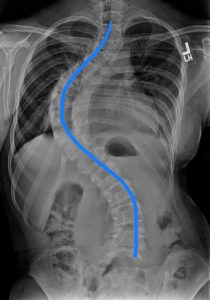
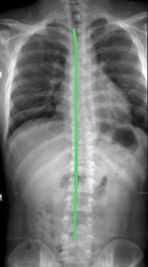
The image with the blue S curve is a patient with scoliosis and the image with the straight green line shows a normal spine.
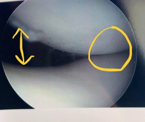
This is a condition where your kneecap is tilted and compresses on the femoral groove abnormally. Imaged below is a picture of a kneecap joint with lateral patellar compression syndrome. You can see where on the right side the bones are touching each other and on the left side there’s a big open space. There should be an even space on both sides.
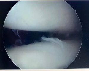
This picture shows how the kneecap looks after I have performed a lateral release. This should give the patient almost immediate pain relief. This is a diagnosis sometimes difficult to figure out because there is not a great imaging modality to document. Many times it is more of a dynamic problem and cannot be statically imaged.
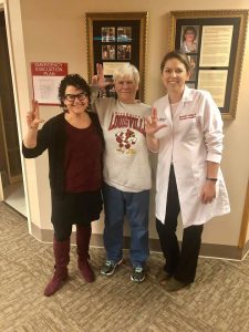
Seeing all kinds of cool orthopaedic stuff! Today we have the privilege of treating one of our University of Louisville Women’s basketball team season ticket holders. She was giving us the scoop on the U of L vs U Conn win. So proud of our lady Cards!!!
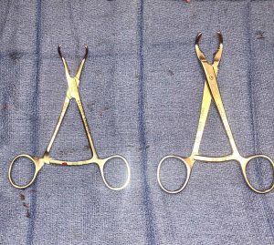
You would think in this day and age of high technology we would have something more technologically advanced to reduce a fracture in the operating room. However, we still use old-fashioned bone reduction clamps to hold the broken bones together while we are fixing the fracture. Imaged below are two different bone reduction clamps that I commonly use. The one on the left is called a point to point and the one on the right is called a lobster claw.
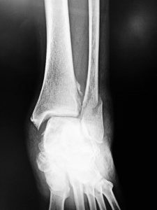
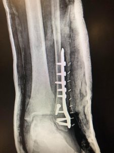
The fibula bone has a spiral fracture and the mortise has been disrupted. A strong stainless steel plate and 11 screws did the trick. Six weeks on crutches, followed by six weeks of PT and lots of home exercises, then this gal will be ready to hit the tennis court again.
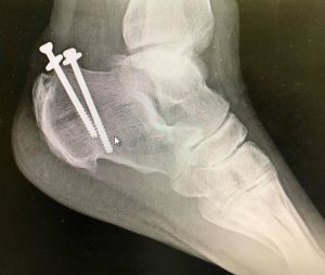
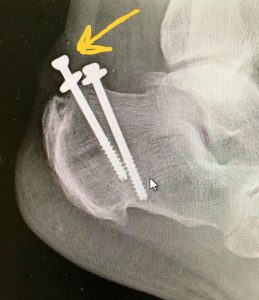
Why do you think the lady imaged below cannot get her shoe to fit anymore ?
And yes, she truly does have a “screw loose” 😜 She presented with the screw head almost pushing through the skin as you can see in the image with the yellow arrow.
The patient had surgery about 15 years ago by another doctor and the screw is just slowly working its way out of the bone. We will have to take that out! 😷


Recent Comments