The skier lost …
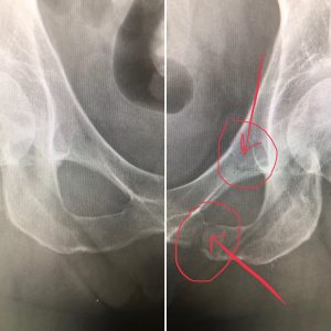
Image on left is a normal pelvis. Image on right with red circle and arrows points to the fracture of the inferior and superior pubic rami.
The skier lost …
Image on left is a normal pelvis. Image on right with red circle and arrows points to the fracture of the inferior and superior pubic rami.
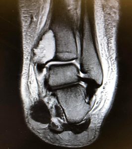
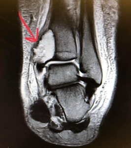
What is that area in the bone that is white, where the red arrow is pointing ?
This patient fell and has acute onset pain involving her ankle. She has a bone cyst also known as a aneurysmal bone cyst. She was most likely born with this cyst and never knew it until she had a trauma. We took an x-ray in the office which showed the cyst and then obtained an MRI scan. The arrow is actually pointing to where she broke through the bone where the cyst is located. This does not appear to be malignant. This will heal with immobilization in a cast boot.
1. Sharp, stabbing pain located in your knee joint.
2. It is easy to localize the pain in the knee and it is consistently in the same place.
3. No pain occurs when resting. Pain occurs with a plant and a twist, lateral motion or flexion of the knee.
4. Slight swelling is present and the knee can feel tight with a slight limit of motion.
5. Clicking, locking of the knee and popping may occur.
Attended a great meeting this morning on girl bosses at the Kentucky Derby Museum with my two amazing hard working and motivated #girlbosses! An amazing panel of women: Tonya Abeln, Raeshanda Johnson, Iris Wilbur and Elizabeth McCall.
My favorite quote of the morning was from Raeshanda, “If somebody closes a door, I go back and buy the building.” Tonya is the director of Community Relations at Churchill Downs. Raeshanda owns and runs her own fashion house: All is Fair in Love and Fashion. Iris is the Director of Government Affairs and Public Policy at Louisville Inc. Elizabeth’s an Assistant Master Distiller at Woodford Reserve. Amazing women!!!


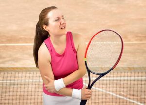
1. Purchase a racket that is flexible. Stiff, high power level rackets will transmit the force the ball produces when it strikes the racket up to the elbow area, irritating the common extensor tendon.
2. Use strings that are poly filament. A monofilament string tends to increase the force on the players elbow.
3. Have your tennis pro string your racket about 2 to 3 pounds lower than normal.
4. Over wrap the grip on your racket by one or two layers. A smaller grip encourages and transmits more tension on the common extensor tendon when holding the racket.
5. Take a lesson from your friendly tennis pro. How many times have you watched a major tennis tournament on television or live and saw one of the pro players wearing a tennis elbow band. You don’t. Because they have perfect technique. Hitting the ball late can contribute to tennis elbow.
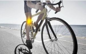
1. Raise your seat slightly by half an inch.
2. Lower your gear so you are pushing an easier gear with higher cadence.
3. Focus on using your hamstrings not just your quads. During your pedal stroke pull back instead of just pushing down.
4. Check your clip-less pedals to make sure they are in a neutral position and not too internally or externally rotated.
5. Consider clip-less pedals that allow for some float. This gives some movement with internal and external rotation.
6. Wear shoes with stiff sole.
7. Consider a bike fit if the above fails. If in Louisville, Kentucky, Curtis Tolson is a certified bike fitter.
1. Pain on the lateral or side of the shoulder is gradual.
2. No significant traumatic event.
3. Pain prevents you from falling asleep at night.
4. Overhead and activity behind the back is difficult and painful.
5. Use of NSAIDS does not help with pain reduction.
1. A pop occurs with the use of the shoulder.
2. After the pop is felt or heard, you are unable to fully lift your shoulder.
3. Night pain that makes it difficult to fall asleep.
4. Pain that is located over the outer side of the shoulder.
5. Activities such as putting your arm behind tour back are difficult and painful.
This is not a good result after a successful dunk of the basketball. Good news is that they were non displaced and short arm casts were applied. What has been your worst broken bone experience?

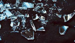
1. Gout is the deposition of uric acid crystals.
2. Pseudo gout is the deposition of calcium pyrophosphate dihydrate crystals.
3. Both conditions produce sharp crystals which are extremely painful. Think of it as throwing a bunch of broken glass into your joint. That’s what happens with gout and pseudogout.
4. Gout may occur with a diet that is rich in meat while Pseudogout has no precipitating factors.
5. Gout is treated with a medication called colchicine for acute events and a medication called allopurinol for long-term preventive treatment. Pseudogout is typically treated with a steroid acutely and there is no long-term prevention medication treatment recommended.
Recent Comments