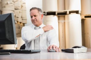
ACL Treated with Hamstring Graft

An ACL Tear Treated with a Hamstring Graft

ACL tear needs to be reconstructed in most patients . People that are active in twisting and turning activities need an ACL https://orthoinfo.aaos.org/en/diseases–conditions/anterior-cruciate-ligament-acl-injuriesWe cannot suture the torn ACL back together. Therefore, the ACL must be reconstructed. I recommend the hamstring graft.  ACL tears reconstructed  with a hamstring graft is excellent. The hamstring graft is A strong graft. In addition, a hamstring ACL autograft , causes less pain at the time of surgery and has a smaller scar.  For example, other graft options include:  the central one third of the patellar tendon or a portion of the quad tendon. Most importantly, an autograft is used for ACL surgery. Similarly, autograft is tissue from your body. Graft selection is surgeon dependent . Because of these reasons, my first choice is the hamstring auto graft for ACL reconstruction surgery https://louisvillebones.com/

ACL Auto Hamstring Grafts are the Strongest

The autografts have the lowest re-rupture rate. In addition, allograft or a cadaver graft has the highest failure rate. Above all, is not recommend to use a cadaver graft for the first time ACL surgery . For instance, revision ACL surgery is commonly done with an allograft or cadaver graft.

Dr. Stacie Grossfeld showing an ACL hamstring graft before it gets fashioned into an ACL auto hamstring graft

Dr. Stacie Grossfeld is showing a hamstring from her patient, that is getting ready to be fashioned into a hamstring ACL graft . Autografts are a the strongest grafts with the lowest rate of re-rupture.

The Hamstring Auto Graft

An ACL tear treated with a hamstring graft gives great results. The hamstring graft has a cosmetic type and  small  incision. Secondly, obtaining the hamstring occurs with a tendon stripper. Thirdly, I then prep the graft on a device called a graft master. The next step involves, suturing  a device that attaches the graft to the femoral side of the ACL. The fixation of the graft to the tibia has many different hardware and device options. The fixation devices are surgeon dependent.

Close up image of an ACL hamstring autograft

Dr. Stacie Grossfeld has just harvested a hamstring graft . This is a close up image of an ACL hamstring autograft .

The surgical procedure involves drilling one tunnel across the tribal and the femur. The surgical procedure takes about an hour to complete. The surgery is outpatient.

Patient of the Month Sherry Hughes

Meet this month’s patient in the spotlight Sherry Hughes!

Sherry Hughes
Originally from Louisville, Sherry has been married for almost 49 years and has two grown and married daughters Ellen and Jane. In her spare time, she enjoys having the opportunity to care for her 4 grandchildren – 3 girls and 1 boy. “I love my role as “Grammy!”
A graduate of the University of Louisville, Sherry followed her father’s footsteps by pursuing a career with South Central Bell. Now retired, Sherry has pursued several part-time jobs over the years, including Pottery Barn Kids and now at Hadley Pottery.
“My passion for collecting all of my dinnerware first began while sitting in the bargain aisles and inspecting the remnants before I got married to very recently working in the retail store a few days a week now.”
Recently, Dr Grossfeld helped Sherry through some painful episodes with her feet after an injury was misdiagnosed by another provider and she was recommended to the Orthopaedic Specialists. Luckily, since her treatment with Dr Grossfeld, Sherry’s discomfort has been kept to a minimum until her most recent shoulder injury.
“This summer on June 1st, I tripped over our dog’s bed out on our deck and took a very ungraceful fall which caused my right shoulder to be dislocated.  I saw the doctor the following day and was given some pain medication and instructions on how to handle this new inconvenience. Since then I have seen Dr Grossfeld who prescribed physical therapy and also gave me a cortisone shot to alleviate the discomfort, especially while sleeping.  All of this has helped and the shoulder is improving!”

Ankle Sprain Treatment Helps Early Recovery

Early Treatment for Ankle Sprains Result in a Quicker Recovery

Early treatment for ankle sprains  results in a quicker recovery. An ankle sprain is an injury that occurs with trauma. In addition, when  the twisting injury occurs, up to 4 major ligaments in the ankle can tear. Therefore it can result in swelling, bruising and  pain.

Mechanism of Injury for an Ankle Sprain

Excessive inversion or eversion motion to the ankle causes a sprain. Inversion is when the ankle turns inward . An eversion  injury occurs when the foot and ankle turn outward. Many times, a pop is heard at the time of injury. After that typically swelling quickly occurs. In addition, it may be difficult to bear weight after the injury.  The amount of pops heard along with the amount of swelling typically correlate that the number of ligaments torn at the time of the injury.

Ankle sprain

Ankle Sprain . Drawing is from the Mayo Clinic


What to Do Next After You Have Sprained Your Ankle?

The first thing to do is place ice on the ankle. The ice must cover the whole ankle joint. Secondly, wrap the ankle with an ace wrap or cobain. If you do not own:  place a kitchen towel around the ankle using safety pins to hold in place. Thirdly, take an NSAID medication , if you do not have a history of stomach ulcers, kidney disease or are taking any blood thinners. If the ankle is too painful to bear weight if might be fractured . If there is concern about a fracture or broken bone, a visit to the orthopaedic surgeon or urgent care is needed https://louisvillebones.com/The provider will examine the ankle and probably take x-rays . The next step is to reduce the swelling and pain.

Treatment Over the Next Six Weeks

Treatment over the next six weeks can be divided into three phases. The first phase is to reduce swelling and control pain. Ice, NSAIDS, ace wrap , sometimes a cast boot , crutches and physical therapy. The quicker PT is started the quicker recovery occurs.

In addition, the second phase is involves weaning out of the cast boot into a lace up ankle brace. For instance the NSAIDS are typically stopped and icing can occur less often. Physical therapy moves to a more functional type of rehab to restore normal activities of daily living. The third and final phase to to introduce normal fitness activities . Therefore the final phase involves more PT https://prorehablou.com/which is sports specific. Therefore, the brace is  worn for another 6 weeks even after PT has stopped. It helps to improve proprioception.

If proper rehab is important so recurrent ankle sprains do not recur  easily .

The Pediatric Wrist Buckle Fracture is Common

Pediatric Wrist Buckle Fracture

The pediatric wrist buckle fracture is a common broken bone pattern in kids. It is a broken bone that commonly involves the radial bone in the wrist. For instance, the fracture occurs from a fall on an outstretched arm. There may be no noticeable deformity of the arm after a buckle fracture occurs. The arm will be painful to touch and swelling typically occurs. In addition the child may not use the affected arm.

Distal radius fracture buckle fracture

A child with a buckle fracture of the distal radius. The red arrow reveals the fracture

What To Do Next

if you suspect your child may have a broken bone as known as a fracture, it needs to be evaluated by a physician. The first thing to do is to immobilize the arm with something. Get creative : a ruler, large serving spoon , and wrap with an ace wrap .  If this occurs over the weekend, stabilize , then head to your local urgent care center . X-rays will be obtained . After that the diagnosis is made. A buckle fracture is easily seen on plain x-rays. The doctor will then split the fracture. The next step will be to get an appointment with an orthopedic surgeon for definitive treatmenthttps://louisvillebones.com/

Treatment by the Orthopaedic Surgeon

The orthopedic surgeon will review the x-rays from the urgent care center. Sometimes repeat films are taken. Above all, the arm will be placed into a cast or waterproof removable cast brace. After that the  follow-up is typically one to two weeks after the visit.

Activity after the Bracing or Casting

If the fracture is a buckle fracture, sporting activities are typically resumed in the cast/ cast brace. In addition , the fracture typically takes four to six weeks to heal. Most kids can resume sporting activities as long as they can play immobilized . Similarly, playing a musical instrument and computer use will resume.

Long Term Effects

Buckle fractures do not cause any long term effects. Healing occurs and then the child will have resumption of all activities .

What are Rotator Cuff Tear Symptoms?

The rotator cuff is a group of four tendons that are located in the shoulder. When a portion of the tendon tears, it can produce pain and disability. This injury is quite common and often requires surgical repair. Here are some of the questions I get asked daily as an orthopaedic surgeon:

Rotator Cuff Tear Symptoms

Above all, the most common symptom of a torn rotator cuff is night pain. Secondly, pain with overhead activity or there is difficulty with bringing you arm behind your back, such as putting your wallet in your back pocket or zipping up a dress.

Where is the Pain From a Torn Rotator Cuff Tear Located?

The pain from a rotator cuff tear is located on the lateral aspect of the arm. Pain is in the area of the deltoid muscle. In addition, the pain radiates down the arm, sometimes to the elbow.

What Causes the Rotator Cuff to Tear?

Most rotator cuff tears occur with age. Therefore, rotator cuff tears tend to be degenerative in nature. Traumatic cuff tears are less common. Traumatic cuff tears are closely associated with shoulder dislocations in people over age 60. In addition, a fall that results in a person landing on a flexed elbow can also traumatically tear the rotator cuff. However, it is rare to tear your rotator cuff if you are under age 40. Most cuff tears occurs in patients 50 years or older, because as we age our rotator cuffs weaken. They can tear with just normal activities of daily living!

Key Findings on Physical Examination of a Rotator Cuff Tear

Key findings on physical examination of a rotator cuff tear is pain located over the lateral side of the upper arm. The doctor will perform physical tests that include a Neer Test and Kennedy-Hawkins Test, as well as placing the shoulder through a range of motion. In addition, strength will be tested, pulses evaluated and sensation tested.

Imaging demonstrating the key examination tests for a rotator cuff tear

The image reveals the key physical tests to perform to document a rotator cuff tear.

Imaging needed to Diagnosis a Rotator Cuff Tear

The initial imaging is a standard shoulder x-ray. If the x-ray does not reveal the source of shoulder pain a MRI will be obtained . A MRI will show soft tissue, bone edema, and detailed areas of arthritis that plain x-rays cannot reveal. However, if the patient has a pacemaker, MRI can not be obtained. In addition, it is difficult for claustrophobic patients to undergo a that study.

MRI of torn rotator cuff

The circle reveals where the rotator cuff tear is located on the MRI.

Treatment of Rotator Cuff Tears

The primary treatment option for a repairable rotator cuff tear is surgery. There are some rotator cuff tears that are not reparable; Massive tears with fatty atrophy and old chronic tears with significant retraction are not amendable to surgery.

Surgery is outpatient and arthroscopic. The surgery is relatively low risk. Smokers put the repair at risk for not healing, as the nicotine in cigarettes inhibits healing. A massive tear greater than four centimeters has a high rate of non-healing. In addition, tears were the rotator cuff is thin, avascular and friable have a difficult time healing even after the perfect surgical reconstruction.

Image of a torn rotator cuff tear at the time of surgery arthroscopically

A torn rotator cuff tear at the time of surgery arthroscopically.

A repaired rotator cuff tendon

A repaired rotator cuff.

Recovery After Rotator Cuff Repair

The recovery after a rotator cuff repair is around six months. However, improvement will continue to occur up to one year after the surgery. The recovery process can be divided into three six-week phases. You will be working with a physical therapist throughout each phase. In the initial six weeks, the patient will not be able to actively raise their arm. Overtime, as you rebuild strength, you will be able to return to a wider range of motion. The last 6 week block of recovery is focused on strengthening. Of course, each person’s recovery process is different, and may vary slightly in length.

If you are seeking rotator cuff treatment and repair in the Louisville, Kentucky-area, board certified sports medicine physician Dr. Stacie Grossfeld at Orthopaedic Specialists PLLC can help. Orthopaedic Specialists PLLC is accepting new patients, and same day/telemedicine appointments are available. For additional information or to schedule an appointment, please call us at 502-212-2663.

Treatment for a Torn Achilles Tendon

Treatment for a torn Achilles’ tendon is typically surgery. Surgery has been showing to increase the patient’s strength compared to non-surgical treatment. The risk of the tendon re-tearing after surgery is less. Patients’ power in the leg is greater when surgery is used to treat the torn Achilles. The non-surgerical treatment has a higher re-rupture rate and less chance of returning back to the strength of the affected leg.

Who’s is a Poor Surgical Candidate for Achilles Tendon Repair Surgery?

Poor surgical conditions for treatment of a torn Achilles’ tendon fall into two groups. These patients are diabetics and morbidity obese patients. Therefore these two groups have a much higher risk of infection and wound complications.  Because of this, as orthopaedic surgeons, we typically recommend treatment in a cast for the diabetic and morbidly obese patient.

What is the Achilles Tendon?

The Achilles’ tendon connects the calf muscle to the heel bone. Secondly, it is a very superficial tendon. In addition, the achilles tendon is easy to palpate and see. Inflammation can occur in the tendon.

Who Tears Their Achilles Tendon?

Most commonly, men (85%) between the ages of 25-45 years old, tear their Achilles’ tendon. This most commonly occurs  while playing sports, as shown in this video. The typical story is the man is playing basketball or tennis. While playing,  they push off with their foot. In addition, they feel a pop. Because of injury, acute onset of pain occurs and its difficult to walk of the court or field  . Within hours the area of the Achilles’ tendon swells.

Achilles Tendon Tear, Best treatment for Achilles Tendon Injury

How the Achilles Tear Diagnosed?

The most common way to diagnose an Achilles’ tendon tear is by physical examination. A defect in the tendon is felt by the healthcare provider. Placing the patient in a prone position makes it easiest to feel. The provider runs their finger down the tendon. There will be a palpable defect in the tendon.  Thompson test is a physical exam To diagnosis an Achilles’ tendon tear.  MRI scan is the best imaging study for documentation. See more info here.

Achilles Tendon Surgery

Achilles Tendon Surgery is performed as an outpatient procedure. The surgical procedure involves repairing the two ends of the torn tendon back together. There are many different techniques. The results are excellent in most patients. It takes 6-8 weeks for the tendon to heal. The rehab after the tendon to heal takes another 2-3 months. Above all, there is a very low re-rupture rate after surgery. Most patients will return back to the same types of sporting activities prior to the Achilles’ tendon tear.

Preventing Overuse Injuries

Overuse injuries. They can stop you in your tracks and prevent you from moving your body the way you want to. Fortunately, there are a few things you can do to mitigate your chances of developing an overuse injury. Read along to find out some general rules you can follow to stay healthy.

Gradual Increases

One of the most common ways people can experience an overuse injury is by taking on too much too fast. It’s all too easy to get in over your head with a new type of exercise or activity. Doing too much of an unfamiliar exercise in a short timeframe puts a lot of stress on the body, in ways it is not adapted to yet. To avoid this, you should always gradually build up your intensity in and time spent on a new activity.

Adequate rest time is an important practice here as well. Your body needs time to recover from hard work, particularly when it is not used to a certain type of work. When you start out with something new, it’s always smart to err on the side of caution and incorporate plenty of rest between workouts. Over time, as your body adapts, you may be able to adjust the amount of rest you take.

Develop Good Form to Prevent Overuse Injuries

Improper form is another way that overuse injuries can occur. If you are moving your body in an inefficient and improper way, it’s easy to overload specific muscles, tendons, and joints. Just like you should gradually build up your workload for a new exercise, you should slowly develop proper form right from the beginning. This way, as you practice more you will develop sustainable movement and help prevent chronic issues from arising.

Get the Right Gear

Depending on the activities you enjoy, you may need more or less specialized equipment to stay healthy. In general, though, the right gear can be a significant factor in your chance of developing an overuse injury. For instance, if you take up cycling and ride on a bike frame that’s too small for a while, you may develop a sore back, knees, and ankles, among other things. Or, if you’re taking up walking or running but do not have well-fitting supportive shoes, you might develop joint pain. Make sure you get the right fit for your activities- it can make a huge difference.

Cross-training is Your Friend

Regularly doing different types of activity that work different parts of your body is key to prevent overuse injuries. Variety is a powerful tool to help you both stay active and healthy. For example, if you love to run, you could consider cross-training with some cycling, core work, weightlifting, and climbing, just to name a few options. It can be tempting to simply put in more time with what you are really focused on. However, that can be a recipe for an overuse injury. When you cross-train, your body uses different muscle groups and is less likely to overload any one area.

If you are injured and you are concerned that you may have an overuse injury, you should seek diagnosis and treatment from a qualified medical professional like Dr. Stacie Grossfeld. To schedule an appointment with Dr. Grossfeld, call Orthopaedic Specialists at 502-212-2663 today.

Injury Heat Versus Ice? What is the Best One to Use?

What is the Best Way to Treat an Injury or Joint Pain: Heat Versus Ice?

why does my knee hurt

What is the best way to treat and injury or joint pain: heat versus ice? Ice versus heat is a common question I get asked as an orthopaedic surgeon on a a regular basis https://louisvillebones.com/Acute injuries that produce swelling are best treated with ice. Arthritis flare ups are best treated with heat. Ice can actually make an arthritic joint hurt more. The cold from the ice will cause the blood vessels to constrict. Therefore it will slow the inflammatory process down right after an injury occurs.

How Long Should the Ice Be Applied ?

There is a myth that ice should only be left on for 20 minutes at a time. Ice can be left on for hours. In other words, the longer it is on the better it will work. The only caution is make sure the ice does not have direct contact with the skin. Because of this,  it can in extreme cases , cause frost bite.

What is the Best Way to Keep Ice on an Injuried Area?

The best way to apply ice to an injured joint is with a motorized ice machine. There are different models on the market https://www.breg.com/products/cold-therapy/devices/cube-cold-therapy/. The ice machine is basically a ice cooler that plugs into the wall. First of all, the ice cooler plugs into the wall, the motor pushes the ice cold water into a rubber tube that attaches to a neoprene water proof wrap. The wrap is placed around the joint. Typically insurance companies will not over the cost of the unit. I highly recommend the ice machines to my patients that have had surgery. The ice machines can give you 9 hours of constant cold therapy. They can help reduce post operative pain medical use.

However, if you prefer not to spend the money on a motorized ice machine there are other options. My favor is a   ziplock bag with ice and water. The water will allow greater contact and molding around the joint. Another good option is a bag of frozen peas or corn. Alway keep extra bags in the freezer for easy and quick trade outs.

When Should Heat Be Used?

What is the Best Way to Treat an Injury or Joint Pain: Heat Versus Ice? Heat typically works best for an osteoarthritic joint that has had a flare. Arthritis is a condition that will have symptoms that wax and wan. It cycles through inflammatory phases and non-inflammatory phases. People with arthritis can have very minimal symptoms when the arthritis is in a non-inflammatory phase. For example, arthritis  can enter an inflammatory phase from a change in weather, activity or an injury. In addition, heat can help reduce the symptoms of arthritis when it is is the inflammatory phase. Heat to the painful joint will increase blood flow. Natural anti-inflammatory factors are located  in the blood stream. Above all, these factors will help reduce the pain, swelling and stiffness.


What is the Best Way to Apply Heat ?

A moist heating pad is a great way to apply heat to a painful joint. A heating pad that emits a moist heat tends to give patients better relief https://www.bestadvisor.com/moist-heating-pads      For instance, if you do not own a heating pad there are homemade options. For example, a moist towel thrown in the microwave then placed in a ziplock bag is a way to apply heat to a painful joint.

Why Does My Knee Hurt?

Why Does My Knee Hurt?

Why does my knee hurt? This is a common question I get asked as an orthopedic surgeon  https://louisvillebones.com. For instance, common causes of knee pain are osteoarthritis, cartilage tears or  ligament injury. Knee pain can start after an injury . In addition, it can also start after overuse.  Knee pain may occur with changes in the barometric pressure.

The ACL is a Common Ligament Torn in Sports Injuries

An injury to the knee can cause a ligament tear https://orthoinfo.aaos.org/en/diseases–conditions/anterior-cruciate-ligament-acl-injuries. The most common sports related ligament that gets injured is the ACL (anterior cruciate ligament). This is the ligament that provides stability to the knee joint. Therefore  it prevents the knee from shifting forward. ACL injuries occur while running with quick change of direction . In addition, another  mechanism of injury can occur with landing from a jump or a sudden stop. Tearing of the ACL causes  a pop felt in the knee . The knee swells within 12 hours of the injury.

The Meniscus is the Most Common Cartilage Torn in the Knee

There are two different meniscus in each knee joint. Each one acts as a shock absorber in the knee. In other words, they help to cushion the joint. The meniscus typically tears from an injury. In contrast, as we age they can tear with normal daily activity and no trauma. A torn meniscus causes a sharp localized pain in the knee joint with twisting activity. In addition, it also cause swelling. Painful range of motion can also be present with a meniscal tear.


Osteoarthritis Can Cause Knee Pain

Osteoarthitis is a process where the cartilage that covers the ends of the bones in a joint breaks down. For instance , some symptoms  of knee arthritis range from morning stiffness, swelling, pain, lose of range of motion, and angular deformities of the knee joint https://orthoinfo.aaos.org/en/diseases–conditions/arthritis-of-the-knee/. Above all, genetic predisposition causes osteoarthritis . Arthritis in the knee occurs from being overweight. The natural aging process causes knee arthritis . Trauma such an intra-articular fracture can cause post -trauma arthritis in the knee joint. The pain quality associated with osteoarthritis tends to be a dull throbbing aching pain that can also occur at night .

An Overview of Radial Tunnel Syndrome

Radial Tunnel Syndrome (RTS) is a chronic condition caused by pressure on the radial nerve. This nerve runs the entire length of the arm, starting in the armpit and running along the outside of the arm to the hand. At the elbow, the nerve goes through what is called the radial tunnel, a thin space made of bone, muscle, and tendon. RTS can be caused by compression on any point along the length of the radial nerve. However, it is named after injury to the radial tunnel itself, where it is especially common.

Symptoms and Causes of Radial Tunnel Syndrome

RTS is often difficult to diagnose due to its similarity in symptoms to more common conditions such as Tennis Elbow and Carpal Tunnel Syndrome. Generally, RTS is characterized by dull, aching pain in the elbow or top of forearm, but can sometimes reach down the forearm and into the back of the hand and wrist. Besides pain, symptoms include weakness, tenderness, and tingling. Patients often have difficulty flexing fingers or rotating the wrist, due to the discomfort. The pain often gets worse at night, too.

Common causes of RTS are repetitive motion and overuse of the arm. Routine pushing, pulling, and twisting can cause RTS, often through sports or physically repetitive jobs like manufacturing. Direct injury such as bone fractures or tissue damage can also cause RTS. Some diseases can make you more susceptible to RTS, such as diabetes or low thyroid.

Treatment and Recovery

If diagnosed with Radial Tunnel Syndrome, there are many forms of treatment available. Treatment is normally based on the cause and the severity of the RTS.

If the RTS is caused by repetitive motions, simply removing those motions from daily life can provide relief. Splinting the arm or wearing a wrist brace to limit motion can also be beneficial. To build strength, physical therapy exercises may be added and continued over time. To reduce discomfort, electrical stimulation, icing, and anti-inflammatory medications are often recommended. Corticosteroid injections are often considered as a non-invasive treatment option to relieve pressure on the radial nerve.

Surgical Option

When the above methods fail to work within 4-6 weeks, surgery is an option. Surgical treatments aim to release the points of pressure on the radial nerve by cutting the compressing tissues, often directly at the radial tunnel itself. A week after surgery, patients can begin recovery exercises and continue physical therapy for several months.

Recovery depends on one’s effort to uphold treatment guidelines and prevent opportunity for re-injury. Additionally, maintaining arm strength and properly warming up before exercise decreases risk of RTS. Reducing repetitive activities long-term can lower the odds of re-injury. Regular exams are also recommended to ensure prevention.

If you are injured and you are concerned that you may have radial tunnel syndrome, you should seek diagnosis and treatment from a qualified medical professional like Dr. Stacie Grossfeld. To schedule an appointment with Dr. Grossfeld, call Orthopaedic Specialists at 502-212-2663 today.