“It’s as easy as riding a bicycle!” This expression has been used to describe tasks that are relatively simple. The fact is, while riding a bike is a great option for leisure, transportation, and exercise, it has become a cause of injury for some. With a growing number of cyclists on the roads, bike safety has become a growing concern.
It’s important to practice safe riding habits every time you bike, like using a hand turn signal and wearing a helmet and other reflective gear. While some accidents are unpreventable, there are ways to minimize your chances at being the victim of a bicycle injury.
Facts Abo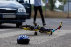 ut Bicycle Accidents – 10 Things to Know
When it comes to safe cycling, there’s plenty to learn. The statistics reported below are not meant to deter riders, but rather to encourage safe practices and caution when taking your wheels for a spin!
- Only 1 percent of all trips are taken on a bicycle in the U.S. (https://1.usa.gov/1fwok7G)
- In 2010, 800 U.S. bicyclists were killed and about 515,000 bicyclists were transported to the emergency room for non-fatal injuries. (https://1.usa.gov/1fwok7G)
- With the amount of injuries and fatalities, biking leads to about five billion dollars in medical and productivity losses. (https://1.usa.gov/1fwok7G)
- The highest rates of bicycle deaths involve 15-24 year olds and adults 45 years and older. (https://1.usa.gov/1fwok7G)
- Males are more likely to be killed riding a bicycle than females. (https://1.usa.gov/1fwok7G)
- In Kentucky, there are laws against riding on the sidewalk and the left side of the street. (https://bit.ly/1TxD2My)
- There are more bicyclist fatalities occur during the summer months, July through September. (https://1.usa.gov/1qHp1yU)
- 21 states including the District of Columbia have laws for helmet use for young riders. (https://bit.ly/1rWW8zk)
- Wearing a helmet reduces the odds of a head injury by 50 percent. Helmets also lower the odds of face and neck injuries by 33 percent. (https://bit.ly/1rWW8zk)
- A biker riding without a helmet is 14 times more likely to die in a bicycle accident. (https://bit.ly/1gjrasd)
 cent over the last 25 years! While the number of marathon runners over age 40 has increased over the last 20-30 years, making 50 percent of all marathons older than 40-years-old.


Recent Comments