
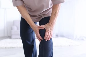
 ACL stands for Anterior Cruciate Ligament and this part of the human body runs between the femur and the tibia. It is one of a pair of cruciate ligaments in the human knee. The other is the Posterior Cruciate Ligament which is located deeper within the knee joint. When an ACL is injured, or torn, it is due to the ligament being overstretched. Most often, ACL injuries occur as a result of a sudden stop or twisting movement. The most common form of repair for a torn ACL is a complete ACL reconstruction. More often than not, the reconstruction is done by autograft and arthroscopically. Despite the repair tissue coming from the same body in which the injury has occurred and the invasiveness of the surgery being minimal, recovery time is set at anywhere between one to two years before a patient can return to normal sports activity.
When Dr. Stacie Grossfeld was training, the idea of performing an ACL reconstruction on a patient over the age of 60 was unheard of. In the past, ACL reconstructions were only offered to patients who were much younger. This was in part due to the extensive recovery time. Over the years, Dr. Grossfeld has found that patients in their 60s who have remained active and show no signs of knee arthritis do benefit from ACL reconstruction. This is especially true in regards to patients that live very active lifestyles. The benefits of undergoing ACL reconstruction are evident when observing patients who are over the age of 60 and have undergone the surgery.
In March of 2017, an article was published in the American Journal of Sports Medicine by authors Dr. Toanen, et al. This group of researchers conducted a study and observed patients who had ACL injuries. All patients were over the age of 60 and athletic with no signs of knee arthritis. These patients underwent ACL reconstruction and their recovery was observed by the group of researchers responsible for the article. What they found was that these patients benefited greatly from the reconstructive surgery. Not only were they able to return to their normal level of activities including: twisting, turning, jumping, and running; they also had no increased incidence of arthritis two years post reconstruction.
Based on these finding, the ground of researchers led by Dr. Toanen concluded that regardless of your age, ACL reconstruction is the best choice for repairing a torn ACL. Age is a factor that should in no way be considered when discussing options for reparation of a torn ACL. Other medical research papers and articles have been published in the past that reinforce Dr. Toanen’s team’s findings.
In December of 2014 a study was done on the long-term outcomes after ACL reconstruction in patients 60 years and older. This team of doctors, led by Dr. Champ L Baker, found that all 13 patients within their study who underwent ACL reconstruction regained their full range of motion and made complete returns to their sports or exercise of choice. This led to the conclusion that “patients aged 60 years or older have good to excellent outcomes with ACL reconstruction and should not be excluded from ACL reconstruction opportunities based on age alone.”
So, Are There Benefits in Undergoing ACL Reconstruction After 60?
In conclusion, yes, there are benefits that come with undergoing ACL reconstruction after the age of 60. Benefits include complete return of mobility, the ability to return to daily sports activity, and the lack of increase in incidence of arthritis. Furthermore, age should not be considered when deciding if a patient is eligible for an ACL reconstruction. If a patient is active, healthy, shows no signs of arthritis in the afflicted knee, and is willing to undergo ACL reconstruction, they should be considered a candidate for the surgery.
If you are 60 years old or over and an active adult who has suffered from a tear in an ACL, contact Dr. Stacie Grossfeld at Orthopaedic Specialists, PLLC in Louisville, KY. Dr. Grossfeld has been routinely performing ACL reconstruction surgeries for over 10 years. She is constantly staying up-to-date on the latest research findings surrounding ACL reconstruction. If you still have questions about ACL reconstruction for patients over the age of 60, call 502-212-2663 and make an appointment today.
 This July, Orthopaedic Specialists is raising awareness about shoulder health through blog posts. These posts will focus on medical research regarding shoulder injuries as well as shoulder strengthening exercises and tips. If you struggle with pain and tightness in your shoulders that prevents you from living your life to the fullest, follow along for five exercises that are clinically proven to increase shoulder strength and benefit shoulder health.
 Lyme disease is a bacterial infection that is transmitted by Ixodes ticks, more commonly known as deer ticks. These nymphal ticks are about the size of poppy seeds, yet they carry the dangerous Lyme disease in their bites. Ticks are thought of as insects, but they are actually arachnids that are related to scorpions and spiders. Deer ticks are found all over the United States and in more than 60 other countries. These ticks attach to one’s skin through questing and are able to feed for several days on one host. If left undisturbed, they are more likely to transmit Lyme disease and other pathogens into one’s bloodstream.
 Coronary Heart Disease (CHD) is the most common type of heart disease to affect Americans and is also the type of heart disease that people are most likely to be talking about when discussing heart disease. CHD occurs when plaque builds up in the coronary arteries. Coronary arteries are the enlarged veins that move blood into the heart. When plaque builds up in these arteries, they narrow and less blood is able to travel to the heart. Narrowed arteries and less blood flow increase the risk of heart attack or chest pain which means it is very important to keep your heart healthy and avoid heart disease.
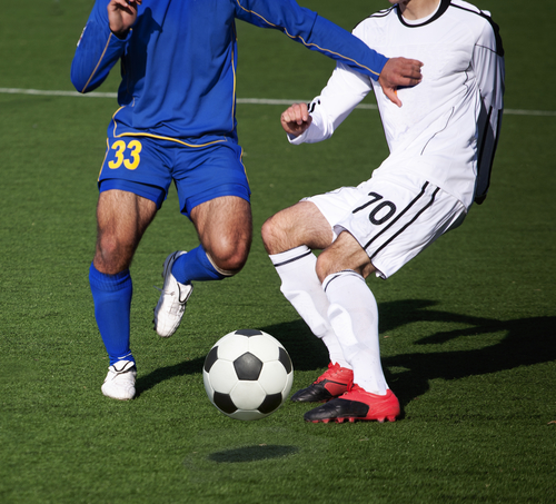

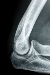 The human elbow is an intricate part of the body. While it is essentially a hinge joint and looks fairly straightforward – the elbow is made up of a complicated series of bones, joints, and ligaments. Within the elbow the radius and ulna must rotate and slide with the humerus to bend or straighten the arm. These movements are possible with the help of ligaments and there are two main ligaments that are important in order to achieve optimal elbow maneuverability: the lateral collateral ligament and the ulnar collateral ligament.
Recent Comments