The Effects of Fitness on the Aging Process
As we age, are there patterns of physical decline? Can those be slowed down or reversed with change in lifestyle? An excellent article was published in the September 2014 volume of the Journal of American Academy of Orthopedic Surgeons. Dr. Bryan Vopat discussed the common physical changes that occur with aging and how to slow down this decline with physical training. A summary of the article is below.
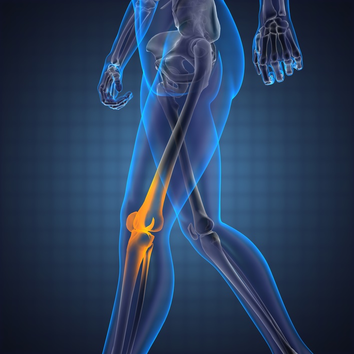
The major changes that occur in the musculoskeletal system with aging are: bone density loss (osteoporosis), the strength and flexibility of muscles decrease (sarcopenia), ligaments and tendons become stiffer and articular cartilage that covers the ends of bones breaks down (osteoarthritis).
Aging Athletes Competing Longer and Performing Better
Evidence over recent years has documented that older athletes are competing longer, and better then they have in the past. They are competing with younger athletes and breaking records within their age groups. These trends are changing the way we think about the older athlete and physical aging.
Older athletes are role models for the concept that age-related decline is not inevitable and that physical activity can counteract the decline in muscle loss, bone loss, and reduced flexibility. More and more athletes are competing in marathon and ironman triathlete events. At the Ironman Triathlon, the number of athletes older than 40-years-old has increased from 25 percent to 50 per cent over the last 25 years! While the number of marathon runners over age 40 has increased over the last 20-30 years, making 50 percent of all marathons older than 40-years-old.
Eight masters (older than age 40) Ironman Triathlon records were set in 2010 with three of the records set in the age group of 65 years and up. For the past 25 years at ironman events, the average performance has improved by 7.5 percent for all age groups older than 45-years-old.
A study that looked at 900,000 marathon runners found that 25 percent are in their mid to late 60s and outperformed half of the runners age 20-54. One of the most interesting findings from the article is the results from the New York City Marathons from 1989 to 2009. This research found that the running times for men older than 64 and women older than 44 years have not yet plateaued. They are constantly improving with the athletes getting faster. This indicates that they have not likely reached their performance limits.
Aging and Bone Loss
Typically women start losing bone mass at age 30 and can lose up to 1 percent per year. After menopause, that percentage rises to 3 percent per year. Men, on the other hand, begin losing bone mass much later. Starting at age 40, men lose bone mass at a slow rate of 0.5 percent per year and that does not accelerate until they reach their 70s and 80s.
Research has shown that exercise can prevent bone loss in the older athlete. A study examining master sprinters, ages ranging from 40-85 years old, maintained tibial bone strength and bone density. Senior Olympic runners over age 65 had significantly greater overall bone mineral density numbers compared to controls. They also found that when comparing the senior Olympic runners to senior Olympic swimmers, the runners had better bone mineral density indicating that weight bearing exercise is critical.
In the seniors who participated in tai chi for 6 months had increased scores (6-9 %) on their bone density DEXA scans, compared to inactive women in their age group. This indicates that some exercise is better than none and that exercise doesn’t have to be performed at a competitive level.
(more…)


 to their last two – one in Vermont and the grand finale in Hawaii.
Recent Comments