Understanding Different Types of Medical Imaging Tests

When your doctor needs to see what’s going on inside your body, they’ll likely order a medical imaging test for you. Medical imaging tests, such as MRIs, X-rays, and CT scans, use different kinds of energy to provide detailed pictures of the inside of your body. In doing so, the imaging tests can help experienced medical professionals diagnose injury and disease, provide treatment options, and monitor existing conditions.

At some point, almost everyone needs some type of medical imaging test. When the time comes, your doctor will provide you with information, but you’ll still likely have questions about the procedure or the differences between the modalities. That’s why we’ve broken down three common tests in the paragraphs below.

An Overview Of 3 Common Medical Imaging Tests

MRI

Magnetic Resonance Imaging (MRI) tests use a magnetic field and computer-generated radio waves to produce high-resolution images of your organs, bones, tissues, and other body structures. A technician will monitor you as you lay down in the MRI machine, which is a long, narrow tube. Usually taking between 30-60 minutes, the process is non-invasive and painless.

MRI tests generally provide more comprehensive, clearer images than CT scans and x-rays, but are also more expensive and time-consuming. Doctors often choose MRIs are to scan the brain, spinal cord, and nervous system, as well as muscles, tendons, and ligaments. Doctors can detect some conditions only with an MRI. These include inflammation, certain illnesses, and types of cancer. Due to the magnetic field, MRIs should not be used by people with metal implants, such as a pacemaker, cochlear implant, or implanted insulin pump.

X-Ray

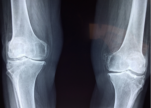
X-ray tests produce images of bones in particular. These tests work by passing x-ray beams through your body that are absorbed in different amounts by different mediums. Denser materials, like bone, show up white, while less dense structures such as muscles and fat show up gray. Compared to MRIs, x-rays are faster, easier, and cheaper—but also may show less detail. X-rays also utilize low-dose radiation, which is not harmful, but can be a concern for some patients.

X-rays are best used to display bone conditions, including arthritis, bone fractures, cavities in teeth, osteoporosis, and bone cancer. Doctors can also use them to diagnose chest conditions such as lung infections, congestive heart failure, and breast cancer.

CT Scan

A computerized tomography (CT) scan takes a sequence of x-ray images around your body, then uses a computer to create a comprehensive cross-sectional image. A CT scan is more detailed than an x-ray, but not as much as an MRI. However, a CT is much faster and more affordable than an MRI. Sometimes it is a good option for victims of car accidents or internal trauma.

CT scans are excellent at showing skeletal system problems, bone mineral density, blood vessels, lung issues, and abdominal abnormalities. They are also good for monitoring tumors and cancer treatment over time.

If you have a condition that you think requires medical imaging, contact your physician for more information. Dr. Stacie Grossfeld at Orthopaedic Specialists PLLC serves people of all ages throughout the Louisville, Kentucky-region in need of all types of orthopedic treatment and sports medicine. For more information or to schedule an appointment, call 502-212-2663 today.

Torn Rotator Cuff: An Overview

Can a torn rotator cuff heal on its own? 

Did you know that over 3 million Americans experience rotator cuff injuries like a torn rotator cuff every year, making it one of the most common points of injury? The rotator cuff is engaged when you use your arm to carry, pick up, push, or bear weight in any way. A group of muscles and four tendons, the rotator cuff’s ball-in-socket structure connects your arm to the ribcage and shoulder, also providing a crucial axis of mobility and stabilization. Needless to say, there’s no way to avoid frequently exerting it in our daily life. 

Rotator cuff injuries range from mild inflammation to a complete tear, and can be caused by several reasons. Based on levels of severity and individual awareness, some people live with rotator cuff tears for months. However, others may seek immediate treatment. Whatever kind of injury you’ve sustained, it’s crucial to know that torn rotator cuffs cannot heal on their own. Not all injuries require surgery, but all require some kind of treatment.  

Common Causes of Rotator Cuff Injuries 

As we age, the natural process of degeneration wears down our muscles and weakens bones, making certain body parts more susceptible to injury. And the rotator cuff, already predisposed to over-use, becomes increasingly vulnerable. Adults over the age of 40 have a steadily growing chance of experiencing rotator cuff injuries. In fact, a reported 30% of adults over the age of 70 complaining about regular shoulder pain. Furthermore, risk factors such as a family history of rotator cuff tears, smoking, long-term repetitive jobs, and bad posture can all increase your risk of rotator cuff deterioration and injury.  

The other primary cause of a rotator cuff injury is through direct trauma, whether it be a fall, pulling on the arm, over-exerting the arm/shoulder, or acutely wounding it in any way. Direct trauma accounts for the more severe spectrum of rotator cuff injuries, and therefore more often requires surgery. Athletes, such as swimmers, weight lifters, and tennis players, are particularly prone to rotator cuff injuries. If you experience this kind of rotator cuff tear, you’ll likely know immediately. Symptoms include sharp pain, a snapping sensation, arm weakness, or popping and clicking sounds.  

Healing and Treatment Options 

Whether you are certain you have a severe rotator cuff tear, or you’re just experiencing mild shoulder pain, it’s highly recommended that you consult your doctor. Untreated rotator cuff injuries only worsen with time. Before your diagnosis, it’s also important that you rest and ice your arm as much as possible.  

Diagnosis will begin with a physical exam by a qualified medical professional like a board-certified orthopedic surgeon. Your doctor may also attempt arm exercises that include reaching forward or rotating the shoulder. After that, if these exercises cause pain, you likely have a rotator cuff injury. Then, based on these results, your doctor may use an MRI, ultrasound, or X-ray to look for tearing. 

If you seem to be suffering from inflammation or only a mild injury, your doctor may recommend using a sling, non-steroidal anti-inflammatory medicine, and/or corticosteroid injections. Then, after immobilization, you will use physical therapy and also personalized exercises to regain your strength and mobility. The majority of rotator cuff injuries require this treatment process.  

Surgery is often necessary for more severe rotator cuff tears. Typically, this is an outpatient, non-invasive procedure. For most tears, surgery will entail arthroscopic repair, in which the orthopedic surgeon will make several small incisions to insert a camera and tools. For more large, complex injuries, the surgeon may choose open tendon repair. Both types of surgery have significant periods of recovery, also requiring months of healing and rehabilitation.  

If you have shoulder pain that is not improving, and are interested in seeking medical attention in the Louisville, Kentucky-region, Orthopaedic Specialists PLLC is here for you. Our experienced team includes double-board certified orthopedic surgeon and sports medicine physician Dr. Stacie Grossfeld. For additional information or to schedule an appointment, call 502-212-2663 today.  

What’s the Difference Between a Rheumatologist and an Orthopedic Physician?

When it comes to seeking diagnosis for chronic pain or an activity-related injury, it’s important to choose the type of medical specialist that’s right for you. Consulting with your primary care physician is the first step. They can provide a diagnosis through x-rays and tests, and, based on your condition, a recommendation for a relevant specialist. But what is the right type of specialist? It can seem like there are a lot of similarities between two different types of doctors, especially if you’re unsure of your affliction. Such is the case for a rheumatologist and an orthopedic physician, both of whom treat the musculoskeletal system. Distinguishing between the two and determining which is right for you can seem daunting.

That’s why we’ve broken it down in this article—keep reading to find out the ins and outs of each.

What is a Rheumatologist?

Rheumatologists are trained to study the musculoskeletal system, including bones, joints, ligaments, nerves, muscles, and tendons. They often treat chronic conditions that affect multiple organ systems, such as autoimmune diseases, inflammatory conditions, or unexplainable musculoskeletal conditions. This includes disorders like rheumatoid arthritis, lupus, scleroderma, or fibromyalgia.

Rheumatologists specialize in pain unrelated to a specific event, or synchronous pain in different parts of the body. Examples can include joint pain coinciding with back pain, reoccurring muscle aches, or any sort of unprompted bone/muscle/joint discomfort.

For treatment, rheumatologists use nonsurgical options, like medicine, physical therapy, or individualized health plans. In the case of most chronic conditions handled by a rheumatologist, there’s no real cure, so they will simply work with you to manage the disease.

What is an Orthopedic Physician?

Orthopedic physicians handle many of the same conditions as rheumatologists, and more. Like rheumatologists, orthopedists specialize in the musculoskeletal system. However, unlike rheumatologists, an orthopedic physician will focus on acute trauma as well as interconnected organ systems.

Orthopedic physicians handle injuries sustained through sports, repetitive activity, or a single incident. A torn ligament, a broken bone, a stress fracture, dislocations—these are all common examples. Locations of specific injuries handled by orthopedic physician include ankles, the back, elbows, hands, hips, the neck, shoulders, knees, feet, and wrists.

Beyond acute injuries, orthopedic physicians can treat chronic or lifestyle-induced disorders like arthritis, osteoporosis, bunions, club foot, carpal tunnel, bone tumors, or hip dysplasia. Long-term damages caused by repetitive movement, like working in a factory or playing tennis, are handled by orthopedic doctors.

While rheumatologists always use nonsurgical treatment options, orthopedic physicians often use surgery in conjunction with nonsurgical treatments. Orthopedists are focused on proactive treatment and future prevention, while rheumatologists typically focus on simply managing chronic illness or inflammation. This means that rheumatologists often refer patients to orthopedists, if surgery becomes a viable option.

One third of all adults in the United States are suffering from some sort of musculoskeletal affliction, at any given time. And it’s no wonder— leading an active lifestyle, working in certain environments, and also just the process of aging itself are all risk-factors. At some point or another, most of us have an injury. What’s important is seeking the right professional for you.

If you are in need of an orthopedic doctor in the Louisville, Kentucky-area who is board certified in orthopedic surgery and sports medicine, contact Dr. Stacie Grossfeld today at Orthopaedic Specialists. We accept most types of insurance and we also offer same day appointments. For more information or to schedule an appointment, call 502-212-2663.

Osteoporosis in Men – 6 Facts You May Not Know

Osteoporosis is a disease marked by a loss of bone mineral density and bone mass. Generally induced by skeletal changes, osteoporosis weakens your bones, putting you at risk of fractures and injuries.

As a man, it’s possible to discount the possibility of experiencing osteoporosis. To begin with, the condition is far more common in women, due to rapid bone-density loss during menopause and women’s smaller skeletal structure. What’s more, osteoporosis is referred to as the “silent disease,” meaning that you often don’t know that you have it until a fracture occurs. With these factors at work, many older men completely disregard osteoporosis, putting themselves in danger. Whether you’re 17 or 70, here are some things about osteoporosis in men that you need to know.

6 Facts About Osteoporosis In Men

1. After the age of 65, men and women lose bone density at the same rate

Although women lose bone-density faster than men during menopause and into their early 60s, by the age of 65-70 the rate is evened out. At this age, your natural absorption of calcium also decreases, putting you at higher risk as well.

2. Health and dietary decisions can greatly reduce the risk and impact of osteoporosis

Exercising and maintaining physical strength, eating a good diet high in calcium and vitamin D, abstaining from excessive drinking, and not smoking can all help limit the chance of osteoporosis. Good sources of calcium and Vitamin D include low fat milk, leafy greens, broccoli, fatty fish, and calcium-fortified foods.

3. Osteoporosis can occur at any age

Although most common in older adults, there are forms of idiopathic juvenile osteoporosis in teens and young adults. The causes of this rare condition are typically unknown, and most sufferers completely recover. That being said, it’s important to be aware that osteoporosis is possible at any age.

4. Your genetic and racial background can impact your chance of experiencing osteoporosis

If you have a family history of osteoporosis or are naturally slender and thin-boned, you may have a higher risk. Also, among men, non-hispanic whites are the most likely to have osteoporosis.

5. Long-term use of certain medications can enhance the risk of osteoporosis

Medications such as glucocorticoids and adrenocorticotropic hormone, antiepileptic medicines, proton pump inhibitors, cancer medications, SSRI’s, and thiazolidinediones can all increase your risk of experiencing osteoporosis or bone loss.

6. You can look-out for symptoms of osteoporosis

If you’re a man older than the age of 65, it’s a good idea to be aware of the early indications of osteoporosis. These include loss of height and weight, changes in posture, gait, and balance, and loss of muscle strength.

Nonetheless, it can be difficult to discern if these are characteristics of usual aging or an underlying condition like osteoporosis. Therefore, it’s always a good idea to consult your doctor—a routine physical exam or blood test can provide insight into your condition.

If you are in the Louisville, Kentucky-area and are in need of an experienced physician specializing in bone health, contact board certified orthopedic surgeon Dr. Stacie Grossfeld for an appointment. Dr. Grossfeld has decades of experience successfully treating patients with osteoporosis. Call 502-212-2663 for more information or to schedule an appointment.

Experiencing Calf Pain? Learn More About Medial Gastrocnemius

If you have a sharp or sudden pain in your calf muscle, you may be experiencing medial gastrocnemius. Commonly referred to as “tennis leg,” medial gastrocnemius is an injury found especially in middle-aged and older athletes. In this condition, there is a tearing of the medial head of the gastrocnemius (calf muscle), which occurs when you overexert or suddenly accelerate. Pain may immediate and sharp, making walking or putting pressure on the leg difficult. Despite initial severity, this injury rarely leads to surgery and can heal in weeks.

Symptoms of Medial Gastrocnemius

Tennis is a highly-competitive and demanding sport, and it’s easy to overestimate your own coordination or turnaround speed, especially as your muscles begin to weaken in middle-age. Although medial gastrocnemius can happen to anyone, it’s most common when you accelerate in irregular or sudden ways, putting undue stress and strain on your body. Tennis leg is also common in high-intensity sports like basketball, soccer, or running.

Being the weakest muscle in the calf, the gastrocnemius is particularly susceptible to injury. In most cases, symptoms are instantly known: intense, piercing pain in the calf. You may feel a sensation of snapping. You may feel like you were kicked or “shot”. There can even be a concurrent popping sound. The injury is tender to the touch, may swell significantly, and may develop some bruising. Walking and moving the leg may seem uncomfortable, and can further agitate the tear. In more severe cases, there may be damage to surrounding tendons. In milder cases or strains, you may only feel pain when using the leg to “push off” in some way.

Treating Medial Gastrocnemius

If you experience medial gastrocnemius, immediate attention is critical to ensure faster recovery. Stopping all activity, immobilizing and/or elevating the leg, and regularly icing the calf can help at first. Consider using crutches, a boot, or even something moderately stabilizing like an ACE wrap. Consultation with an orthopedic specialist like board certified orthopedic surgeon and sports medicine physician Dr. Stacie Grossfeld is crucial, as soon as possible. Doctors will most likely make a diagnosis through an MRI or ultrasound.

Upon diagnosis, your doctor can establish a treatment plan. For medial gastrocnemius, there are three degrees of injury: first degree (stretch injury), second degree (slight tear), and third degree (total rupture). The determined degree of severity will shape your recovery length and process.

In most first and second-degree cases of medial gastrocnemius, activity can be gradually introduced through physical therapy in a matter of weeks. Some physical therapy activities can include passive and dynamic stretching, massage, ultrasound, and rehabilitative exercises. For third degree cases, surgery or more long-term treatment may be required.

If you are struggling with symptoms of jumper’s knee, Orthopaedic Specialists can help! To schedule an appointment with Dr. Grossfeld and the other experts at Orthopaedic Specialists, call 502-212-2663 today.

Patient of the Month Trey Mattingly

Meet this month’s patient in the spotlight Trey Mattingly!

Currently, Trey is a junior in High School and plays baseball for the South Spencer Rebels. However, in the fall he tore his ACL playing football.

“During our second varsity game of the season, I ran down the field on kickoff and ran over a kid trying to get the receiver. I was reaching for the receiver when a kid grabbed me, and I twisted and heard a loud pop.”

“Luckily, Dr. Grossfield was great. We were skeptical that I would not be ready for spring baseball season but I knew with hard work, dedication and a great surgeon I would be ready. And we did it! I recovered months earlier than predicted. I put on about 15 pounds of muscle, and am stronger, faster, and more determined than ever.”

Trey has always been very athletic and was even in a T-ball league at the age of 2!

“I was 2 years younger than most of the kids but I was tall and I knew how to hit and run better than all of them. I loved baseball so much I use to sleep with my glove.”
“I’ve always been very competitive. I love some great competition though because that’s what makes you better. If someone is better than me I work harder than them to better myself. Playing baseball you make tons of friends and I think of my team like family.”

Presently Trey plays left field, 3rd, as well as pitches from time to time.

“I can play pretty much any position. But I like left field the most. I work hard and I’m determined to get to the next level.”

What is a Growth Plate Fracture?

How does a bone get longer? Growth plates. They are responsible for bone growth. Children can experience a growth plate fracture with a traumatic injury. Read along to learn more.

Growth Plates

Each long bone in the body has a growth plate. The growth plate is the area where bones get longer. After children reach skeletal maturity, their growth plates close and no more vertical growth occurs.

The growth plate itself is made up of an area of cartilage with rapid turnover. This area turns cartilage into bone and allows the bone to elongate. Most long bones in the body have two growth plates. There’s one plate at the top of the bone and one at the bottom. Long bones like the femur (the thigh bone), the humerus (the upper bone in the arm), and the tibia (the shin bone) all have two growth plates.

However, growth rates aren’t equal in all areas of the body. For instance, the greatest area of growth in the leg occurs through the the growth plate in the femur closest to the knee joint. Growth plates also close earlier or later in different parts of the body. For example, one of the last growth plates to close is in the collar bone, while the first to close is in the elbow.

Additionally, girls’ growth plates tend to close around age 12-14 years of age while boys’ growth plates close between 16-17 years of age. The hormones that are primarily responsible for signalling growth plate closure differ here. Estrogen is the signal for girls, while testosterone is the signal for boys.

A Growth Plate Fracture

A growth plate can be injured from a traumatic event. Trauma can cause a fracture to go through the growth plate, causing it to partially or completely close prematurely. If part of the growth plate partially closes due to trauma, the remaining portion of the bone can grow crooked. If the growth plate completely closes early, the bone can actually end up being shorter than the corresponding bone on the other side of the body.

It’s not just trauma that can impede a growth plate. Repetitive use injuries can also cause damage to growth plates. Typically, though, this type of injury does not cause permanent damage to a growth plate.

If you experience a growth plate fracture, you should see a doctor. It is very important to make sure the growth plate is lined up in an anatomical position. This will improve the chances that the growth plate will not prematurely close from the fracture.

If you are struggling with a growth plate injury, Orthopaedic Specialists can help! To schedule an appointment with Dr. Grossfeld and the other experts at Orthopaedic Specialists, call 502-212-2663 today.

What is an AC Joint Dislocation?

The AC joint is located in the shoulder where two bones meet. You can experience an AC joint dislocation with some traumatic injuries. Read on to learn more.

AC Joint

The AC joint is located between the clavicle and acromion in your shoulder. AC stands for acromioclavicular (acromion plus clavicle). The joint has a few main parts. First, there is a capsule that helps to hold the joint together. Additionally, there are several different ligaments.

How Does the Joint Dislocate?

The AC joint can dislocate if you experience a direct fall on your shoulder. This commonly occurs from crashing on a bike, falling while skiing, or falling while skateboarding.

Immediately after the trauma, you feel pain over the AC joint. It becomes difficult to raise the arm after the injury occurs. The area may also show signs of bruising. There may be a slight or large elevation of the joint area after the injury as well.

Treating a Dislocation

The first line treatment is to apply ice to the affected area. Ice can be kept on your shoulder as long as you can tolerate it. Initially, a sling may help to immobilize the arm too. This will help to reduce the pain because it stabilizes the injured joint. You may also want to consider anti-inflammatory medications to help reduce pain. Of course, make sure your doctor approves any medications first.

You may find that sleep is difficult with this injury. Sleeping in an upright position may be most comfortable, as it is very difficult to sleep on your injured side. In fact, it may take up to eight weeks before you can sleep comfortably in a normal position.

How Your Doctors can Help

When you visit your doctor for this injury, they will likely perform an X-ray on your shoulder first. Depending on your pain level, they may order a prescription NSAID for you to take as well. Once your pain and swelling is reduced, they may direct you to a physical therapist. Physical therapists can help restore your range of motion of the shoulder . They will also help you rebuild the muscles around your shoulder joint.

Prognosis and Return to Activities

Most people have symptoms for a minimum of six weeks after this injury. Some may experience symptoms for up to six months. It is also not uncommon for joint popping to occur after the injury. Fortunately, surgery is rarely needed for this injury. You can also return to any sports once the range of motion in your shoulder is restored and your pain has diminished.

If you are struggling with knee arthritis, Orthopaedic Specialists can help! We offer various treatment options to help you manage your arthritis. To schedule an appointment with Dr. Grossfeld and the other experts at Orthopaedic Specialists, call 502-212-2663 today.

Diagnosing and Treating Ankle Injuries

If you have experienced a pop around your ankle, it can be pretty unsettling. Today we’re going to go over two common ankle injuries that can cause this: an Achille’s tendon tear and an ankle sprain. Read along to learn more about these two ankle injuries.

Achilles Tendon Tear

Achilles tendon tears are most likely to occur in men over age 35. The most common injury scenario for this injury involves playing basketball, tennis or another sport that requires significant pushing off with the ankle. At the time of the injury, a person with a tear will feel a large pop. Most people also describe a feeling like someone kicked them in the heel when the injury occurs. Immediately after the injury, there  is swelling and pain, and it becomes difficult to walk.

Doctors can diagnose an Achilles tendon tear with a physical exam in which they feel for a defect in the tendon. They may also use an MRI scan to visualize the tear. Often times, though, the diagnosis can be made by a physical examination alone.

Doctors will treat this injury with surgery unless the patient is morbidly obese or diabetic. This is because morbidly obese patients and diabetics are at a higher risk for an infection with surgical treatment. Nonsurgical treatments for this condition involve casting the ankle.

The surgery is an outpatient procedure. After surgery, patients are non-weight bearing for several weeks. After that, they may be cleared for touch toe weight bearing (using toes for balance but not weight bearing). Full weight bearing is usually possible starting around six weeks after surgery. The speed of recovery is based on the repair quality, and recovery differs between patients.

Ankle Sprain

A pop felt on the outer side of the ankle after a twisting injury is often a sign of an ankle sprain. This injury may occur if your ankle rolls as you are putting weight on it. A ligament in the ankle is injured in an ankle sprain.

Often, after an ankle sprain people experience swelling and the ankle turns black and blue. Sometimes the discoloration can travel down to the foot. Doctors typically recommend an X-ray to make sure there are no fractures around the ankle joint.

To treat an ankle sprain, follow the acronym RICE. This stands for rest, ice, compression and elevation, which should be the first lines of treatment. You may also need to use anti-inflammatory medication to help reduce swelling and pain. Long-term, physical therapy is a key part of recovery to restore range of motion and help reduce swelling. Physical therapists can help you regain strength and stability around your sprained ankle.

If you play sports, your return time frame depends on the severity of the sprain. It can be as quick as 2 weeks with a mild sprain that you treat properly. More severe sprains may take as long as 8 weeks for a full recovery. In all cases, you may experience intermittent swelling for up to three months after the injury.

If you have experienced an ankle injury, you can trust the professionals at Orthopaedic Specialists to help you recover. To schedule an appointment with Dr. Grossfeld and the experts at Orthopaedic Specialists, call 502-212-2663 today.

Three Common Knee Injuries

Knee Injuries

You’re planting your leg to make a quick turn and- pop! You hear a popping sound from your knee. This is an all too familiar of a situation for many people. There are three common injuries that cause a pop in the knee. These are an ACL tear, a meniscal tear, and a patella dislocation. Most often, for all of these knee injuries, you will experience pain and swelling. Here’s an overview of these three knee injuries.

ACL Injury

The ACL (Anterior Cruciate Ligament) stabilizes the knee.  Planting and twistin, like while playing soccer or football, can cause an ACL tear.  Running and coming to a sudden stop can also tear the ACL. Regardless of the cause, with an ACL tear, a knee pop and swelling will occur. There’s a significant amount of pain right when the tear occurs as well. In sporting contexts, players with an ACL tear are unable to continue competing due to the severity of the injury. With the proper initial treatment of resting, icing, and compressing, the pain can resolve in a matter of days.

Doctors diagnose an ACL tear with a physical examination. They check your injured knee for swelling and pain, comparing it to your healthy knee. They may also move your knee in a few positions to assess its range of motion. Doctors may also utilize diagnostic imaging like a MRI scan to assess the degree of tissue damage and the severity of the tear. In many cases, particularly for people with very active lifestyles, surgery is necessary to repair the ACL. During surgery, doctors reconstruct the torn ligament You will also need to pursue knee rehabilitation for a period of time to restore function and stability.

Meniscal Tears

Another common knee injury is a meniscal tear. Meniscal tears often occur due to trauma like aggressive pivoting in younger patients. In contrast, older patients can tear their meniscus with minimal force from day-to-day movement. In both cases, the tear causes a sharp pain along the joint line. This pain occurs with activity. A meniscal tear needs to be treated properly- the meniscus functions as a shock absorber in the knee.

Doctors diagnose meniscal tears with a physical examination in which they move your knee in different positions to pinpoint the injury. They often use an MRI to visualize the injury as well. Meniscal tears, like ACL tears, should be treated with rest, ice, and compression initially. Longer-term, treatment includes physical therapy to improve motion and stability, and may also include surgery. In surgery, doctors can repair the tear with a low-risk outpatient procedure. Recovery from this surgery is quick.

Patella Dislocations

One final common knee injury that causes popping is a patella dislocation. The patella, also known as the knee cap, can dislocate when you plant and twist your let. Patella dislocations occur most commonly in younger females. People may feels something slip out of the joint if they experience a dislocation. Fortunately, most of the time, the dislocated knee cap spontaneously relocates on its own. However, on occasion it doesn’t. If this is the case, an ER physician will have to relocate the patella. Relocation is perform under a twilight type anesthetic.

Treatment for this injury involves a patella stabilization brace. Physical therapy is also needed to strength the muscles around the knee cap. Typically, people are able to resume full activity within eight weeks of the initial injury. If the patella dislocates again, you may need surgery. During surgery, doctors can reconstruct the medial patellofemoral ligament, the ligament that stabilizes the patella.

If you have experienced any knee injuries, you can trust the professionals at Orthopaedic Specialists to help you recover. To schedule an appointment with Dr. Grossfeld and the experts at Orthopaedic Specialists, call 502-212-2663 today.